


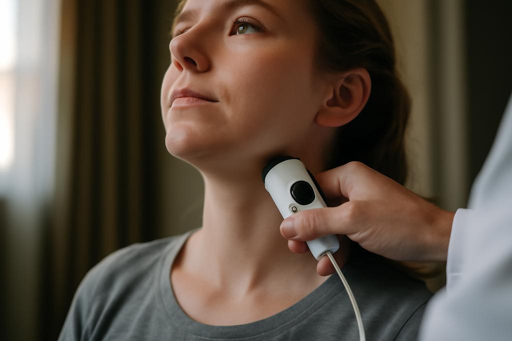
Blood keeps the brain awake, but watching it move without poking the skin is harder than it sounds. The neck, where the carotid arteries ferry oxygen and the internal jugular veins carry away used blood, sits at a crossroads of circulation. In that compact corridor, a new kind of optical listening device is learning to hear the rhythm of flow—like a stethoscope for blood that hums through the neck’s big vessels.
Researchers from Toronto Metropolitan University in Canada and Kagoshima University in Japan, with collaborators at iBEST and St. Michael’s Hospital, set out to test whether simple physiological maneuvers could modulate neck blood flow in realistic, real-world conditions. Led by Gennadi Saiko, and with coauthors Timothy Burton, Faraz Sadrzadeh-Afsharazar, Shota Yamashita, Kenshin Shimono, Yasuyuki Kakihana, and Alexandre Douplik, the team used two kinds of photoplethysmography sensors to peek at changes in the neck’s blood movements while a seven-minute protocol played out. This is not just a tech demo: it’s a deliberate step toward bedside tests that could track how blood travels to and from the brain during emergencies or critical illness.
A new toolbox for neck blood flow
On one side of the neck, a standard clinical PPG sensor sits over the internal jugular vein. On the other, an in-house PPG device built around an Arduino, two LEDs at 660 and 850 nanometers, and a photodiode logs data at 400 samples per second. The two sensors speak the same language in different dialects, and together they probe both the transient pulsations and the slow drift of blood in the neck’s major vessels.
Seven maneuvers were stitched into a one‑minute sequence: abdominojugular testing, breath holding, the Valsalva maneuver, and four occlusions of the internal jugular vein—two on the right and two on the left, proximal and distal. Each maneuver began with a baseline snapshot, then a 15‑second window of active manipulation, then a 30‑second recovery. The goal wasn’t to torture volunteers but to coax measurable changes in blood flow that optical sensors could capture in a setting closer to a hospital ward than a laboratory bench.
Two signals were of particular interest: the AC component, which tracks the pulsing part of the heartbeat, and the DC component, which reflects the overall blood volume in the vessel. The clinical device and the homebuilt sensor both showed that the maneuvers altered the rhythms in the neck, but in somewhat different ways. The Valsalva maneuver—getting a forced breath with a closed throat—stood out as producing the strongest, most reproducible modulation of the neck’s signal.
The study also turned a careful eye to how the signals rise and fall as the heart, lungs, and veins adjust to stress. The right IJV occlusions produced decreases in the AC amplitude on the side of the occlusion, as one might expect when a major drainage path is pinched. But the DC component—the slow shift in the average blood present—behaved more unevenly, hinting at a complex web of alternate drainage routes and venous redundancies that the authors say demand further digging with larger samples and complementary imaging.
Why this matters for medicine and bedside care
Noninvasive, portable, and relatively inexpensive optical tools that can map how blood moves through the neck could become a useful complement to ultrasound and MRI in the ICU or during emergency triage. The brain is an ally with fragile arteries and a sensitive venous system, and tiny shifts in flow can signal trouble long before symptoms appear. If clinicians could watch those shifts in real time with a skin‑hugging sensor, it might help spot dangerous pressure changes, guide treatments, or monitor how a patient responds to a therapy.
The authors argue that contact PPG—a sensor that stays on the skin and can ride along with a patient during care—offers practical advantages in busy settings where noncontact imaging can be hampered by motion or glare. A small, flexible sensor that can slide with the skin could reduce false alarms caused by movement, while a sensor enclosure can shield the detector from ambient light. The flip side is that real rooms aren’t as clean as labs: patients move, lights flicker, and their bodies are different in every way. This study takes a small but deliberate swing at that challenge by testing maneuvers designed to evoke clearer signals.
Yet the work is careful to frame its findings as a first step. With only two healthy volunteers, the team cannot claim to map disease states or to replace established imaging. Instead, the value lies in showing that a compact, dual-sensor approach can pick up meaningful changes when the body is coaxed to redistribute blood in the neck. The next steps, they note, include a larger cohort, cross‑validation with ultrasound, and experiments in conditions more representative of the bedside—perhaps with patients who have head injuries, stroke risk, or congestive heart failure.
What surprised the researchers and what comes next
One of the standout observations was the Valsalva maneuver’s grip on the signals. When people perform this forced expiration against a closed glottis, venous return to the heart is dumped, and the body’s reflexes push back with changes in heart rate and vascular tone. In the neck, that translates into a pronounced drop in pulsatile amplitude and a dropout in the DC level on the in-house sensor. It’s not a clean, one-note effect, but it is repeatable—precisely the trait you want in a diagnostic signal.
Internal jugular vein occlusions tell a more nuanced story. Blocking blood flow on one side reduces the pulsatile swing on that same side, but the slow, baseline signal can swing in unpredictable ways because the brain drains through many channels. The researchers point to the redundancy of cerebral venous drainage, through superficial and deep channels and even extra-cranial routes, as a plausible reason why the DC changes are less consistent than the AC changes. In other words, the neck’s plumbing is messier than a single pipe, and the sensor is reading a chorus, not a solo.
Looking ahead, the team envisions a toolkit rather than a single device: more volunteers, more maneuvers, and more careful synchronization across data streams. They also want to compare their sensor readings with ultrasound—the current gold standard for vein and vessel assessment—and to extend testing into clinical contexts that resemble real emergencies rather than controlled poses. If successful, that toolkit could someday ride in a bedside monitor, whispering when blood flow is shifting in ways that demand attention.


