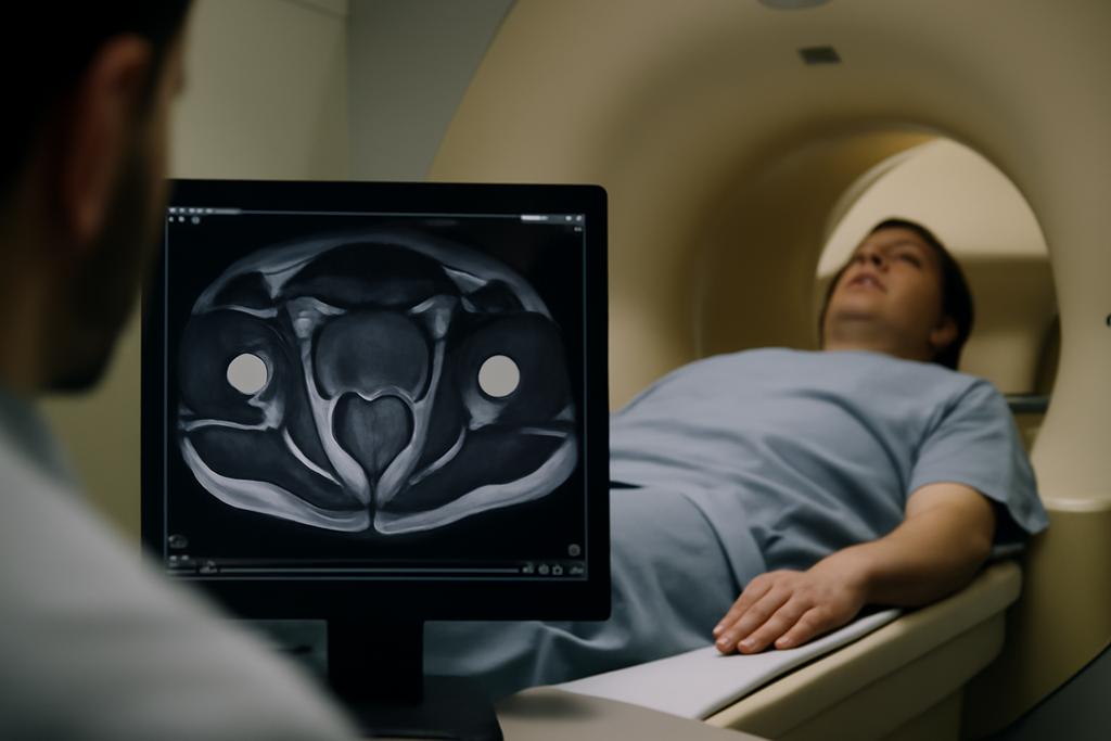
Rewriting the Rules of Deep Tissue Imaging
For years, doctors have struggled to get clear images of deep-seated organs like the prostate using ultra-high-field (UHF) MRI, specifically at 7 Tesla. The problem? At these incredibly powerful magnetic fields, the radio waves used to create the images behave erratically within the body, producing blurry, unreliable results. It’s like trying to focus a camera lens through a dense fog; the image is distorted and hard to interpret. A team of researchers at the University of Duisburg-Essen, New York University Grossman School of Medicine, UMC Utrecht, and the Max Planck Institute for Biological Cybernetics, led by Kristina Popova, Jan Taro Svejda, and others, have discovered a clever solution that could finally change the game.
The Leaky-Wave Antenna: A Novel Approach
Their innovation lies in a new type of antenna, called a leaky-wave antenna (LWA). Traditional MRI antennas, often shaped like dipoles or loops, rely on ‘standing waves’—think of them as vibrations trapped within a guitar string. These create a consistent but sometimes limited field. The new LWA design, however, generates a ‘travelling wave,’ more like a wave rolling across the ocean. This fundamental difference in wave behavior offers unprecedented control over the radio-frequency field.
The researchers’ genius isn’t just about the type of wave, but how the wave is shaped. They’ve optimized the antenna’s design to create a non-uniform phase distribution of the current, a concept they explain as the “phase of currents” along the coil’s conductors. Instead of a uniform, predictable phase, they’ve produced a wave that gradually shifts its phase, precisely controlling how the radio waves interact with the body. This ability to manipulate the wave’s phase allows for better focusing of the radio waves on the target organ, creating sharper, clearer images even through thick tissue.
Focusing the Beam: Mimicking Nature’s Precision
To understand their approach, picture a searchlight. A standard searchlight casts a broad, diffuse beam. The LWA, with its meticulously controlled phase, is like a highly focused laser pointer; the energy is concentrated in a much smaller, more precise area. The researchers achieved this by meticulously designing the antenna’s slots and currents to focus the radio waves deep into the body, precisely at the desired organ, essentially overcoming the challenges posed by UHF MRI.
Their simulations and experiments, using a tissue-mimicking phantom, revealed a remarkable improvement in image clarity at a depth of 7 centimeters—a region relevant to prostate imaging. They found that their LWA generated a significantly stronger radio-frequency magnetic field (B1+) in the target area compared to traditional dipole antennas, improving the signal-to-noise ratio and resulting in substantially clearer images. In short, the improved focus allows for visualization of otherwise hidden details.
Beyond the Prostate: Broadening the Scope
While the study focused on prostate imaging, the implications extend far beyond. This breakthrough could significantly improve the diagnostic capabilities of UHF MRI for various deep-seated organs, potentially leading to earlier and more accurate diagnoses of conditions such as cancer. Imagine a world where detecting early-stage tumors is more reliable and less invasive, leading to improved patient outcomes. The LWA is not merely an improvement, but a potential paradigm shift in the way deep tissue is imaged.
The Challenges Ahead: Scaling Up for Clinical Use
While the results are exceptionally promising, several hurdles remain. This research was conducted in a controlled environment using tissue-mimicking phantoms. Translating these findings to real-world clinical settings will require further refinement, including the development of multi-channel versions for full body imaging and addressing the complexities of human anatomy. The scientists themselves acknowledge the need to scale up the antenna design for clinical applications; they have already indicated plans to create an 8-channel transceiver body RF coil based on their new technology.
The development of this advanced LWA represents a significant step forward in UHF MRI technology. The innovation lies in the meticulously controlled phase distribution of the radio waves, making the LWA a highly focused tool for deep tissue imaging. It’s a technology with the potential to revolutionize how we visualize and treat a wide range of medical conditions.


