


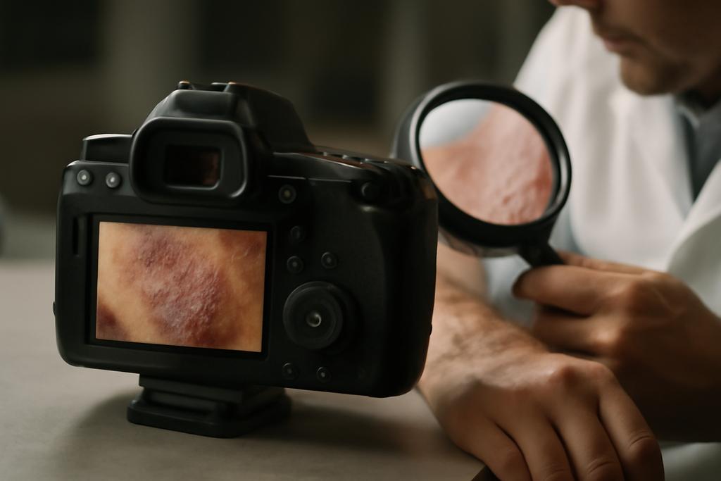
Seeing skin through a cleaner lens
In medical image analysis, the biggest bottleneck isn’t always the brainy guts of a neural network but the first impression the image makes on it. The colors, the contrast, the texture—these are the filters through which a model learns to decide healthy from diseased skin. A team spanning the European University of Bangladesh, BRAC University, and the University of Maryland, Baltimore County, set out to test a simple idea with big consequences: does the way we pre-process skin images change the outcome of the diagnosis? The study’s lead author is Enam Ahmed Taufik, with collaborators across the three institutions, and their goal was to see if tiny tweaks in input preparation could make dermatology AI more accurate and more trustworthy.
What they found isn’t just a marginal gain in accuracy. the pre-processing choice matters a lot, and the story gets even more interesting when you pair it with modern model architectures that can capture complex visual patterns. The researchers compared classic convolutional networks with newer vision transformers, while feeding the same skin-image data through different pre-processing pipelines. The headline result: a particular contrast-enhancement technique paired with a transformer backbone consistently yielded the highest performance and clearer, more localized explanations of the model’s decisions.
Beyond the numbers, the work speaks to a larger truth about AI for medicine: reliability and interpretability hinge as much on data handling as on algorithmic finesse. The study also highlights real-world constraints, like how some pre-processing steps might be cheaper to run in a busy clinic or in low-resource settings, which could determine which systems actually see the light of day. In short, the researchers remind us that the “how you show the data” step deserves as much curiosity as the “how you build the brain” step.
The pre-processing battleground: RGB, CMY, and CLAHE
The team designed a careful, apples-to-apples comparison among four variants of the same skin-image dataset: standard RGB, augmented RGB, CMY color-space transformation, and CLAHE—Contrast Limited Adaptive Histogram Equalization. They curated the dataset by fusing three public sources to cover a broad spectrum of conditions, then addressed class imbalance with targeted augmentation so rarer diseases didn’t get drowned out by more common ones. The result was a robust 7,200-image training pool drawn from 3,468 curated samples spanning ten classes, including several common viral infections and non-musp skin conditions.
In practice, CMY offered sharper boundary cues. the subtractive color space helped delineate lesion edges from surrounding skin, potentially aiding a viewer (and a model) in separating subtle differences. Yet, when it came to learning from these images, most transfer-learning CNN backbones still carried the bias of their RGB-trained origins. The color-space shift proved challenging for these networks, which were pretrained on RGB data. The takeaway: what your model learned during pretraining can constrain how well it adapts to a different color representation.
By contrast, transformer-based architectures—networks that look at an image as a collection of patches and weigh their relationships through self-attention—showed remarkable robustness across pre-processing choices. The same RGB, CMY, and CLAHE inputs yielded surprisingly similar accuracy and F1 scores for these models. In other words, transformers can tolerate shifts in color statistics better than traditional CNNs, likely because their global attention cast a wider net across the image rather than relying on a fixed, localized set of features.
CLAHE, a local contrast enhancement technique, emerged as a particularly practical win. It boosted the model’s ability to distinguish subtle lesion boundaries without the heavy computational burden of some advanced image enhancements. The study notes that while CLAHE improves local contrast, CMY’s linear, low-cost transformation can offer comparable gains in boundary clarity—an important consideration for deployment where compute and power are at a premium. The researchers even point to a potential hybrid path: use CMY when hardware is constrained, but lean into CLAHE when the extra cycles are available for a bit more polish.
Who wins? CNNs versus transformers in the skin-vision arena
When the dust settled, the results lined up with a growing consensus in computer vision: transformer-based models often outperform traditional convolutional networks on complex, high-variability image tasks. Across RGB, CMY, and CLAHE inputs, a large transformer-based backbone achieved the top scores, with accuracy and F1-scores hovering around 0.93 for RGB inputs and staying robust across the other pre-processing options. The best-performing setup didn’t rely on a single trick; it combined the global perspective of a transformer with input pre-processing that sharpened contrast where it mattered most for clinical cues.
Even more telling was the contrast with unprocessed images. CNNs struggled when fed raw input, signaling how sensitive some architectures can be to input quality. The visual language of skin lesions—edges, textures, color shifts—gives CNNs a hard time when the image isn’t perfectly prepped. Transformers, meanwhile, could infer relationships across distant regions of the image, compensating for some noise and variation. The practical implication is clear: if you’re aiming for a robust diagnostic aid in real-world clinics, you’ll want architectures that can reason about long-range structure in images, not just local patches.
Within the transformer family, the study found that some variants nearly matched one another on RGB and CLAHE inputs, with Swin Transformer approaching top-tier performance and a large, modern backbone maintaining a slight edge. The authors emphasize that the improvements aren’t just about a bigger model; they reflect a qualitative shift in how the model processes information—more holistic, less brittle to hue, brightness, or texture quirks that commonly appear in skin images gathered across devices and environments.
In a twist that matters for deployment, the results hint that CMY could be an attractive input alternative in settings where RGB pretraining data are scarce or hardware is constrained. The key is recognizing when the architecture itself can compensate for color-space differences and when it cannot. The paper’s nuanced take is that there isn’t a single universal recipe; the best path depends on the balance of hardware, data quality, and the chosen model family. This insight matters because it reframes the engineering question from “which model is best?” to “which combination of model and preprocessing suits the real-world workflow you’re trying to enable?”
Seeing the model see: Grad-CAM and the heat of trust
A critical part of medical AI is explaining why a prediction is made. The researchers used Grad-CAM, a visualization technique that highlights the parts of an image most responsible for the prediction. They applied it across the four input variants and found that models trained on CLAHE-enhanced images tended to focus more precisely on the lesion regions that clinicians look at when diagnosing. In short, the combination of CLAHE preprocessing with the transformer backbone not only boosted accuracy but also improved the alignment between the model’s attention and human clinical reasoning.
The heatmaps aren’t just pretty pictures. They offer a sanity check for doctors and patients, helping to illuminate when a model is paying attention to clinically relevant features versus wandering into background noise. This interpretability matters as much as the numbers: it builds trust, and trust is a prerequisite for adoption in clinical settings. The authors’ use of Grad-CAM across multiple preprocessing strategies demonstrates a practical pathway to transparent AI in dermatology, rather than a mysterious, inscrutable black box.
Interpretability, in this view, is a tool for safety. when clinicians can see where the model is looking, they can spot when a system might be fixating on irrelevant artifacts or imaging quirks. The combination of robust performance with tangible explanations helps bridge the gap between high-tech accuracy and human judgment—an essential balance in patient care.
What this means for clinics, patients, and the broader AI dialogue
The paper’s takeaways reach beyond the lab bench. First, pre-processing isn’t a mere tidy-up step; it actively shapes what a model can learn. A simple adjustment to image contrast or color space can swing the balance between a correct diagnosis and a missed one. That’s a powerful reminder for developers and health systems choosing AI tools: data handling decisions matter almost as much as the core model design.
Second, the study underscores a practical path toward robust, explainable dermatology AI that can function in a range of environments. CLAHE’s local contrast enhancement, combined with transformer architectures that can ingest information across an image, yields a system that performs well and offers clinician-friendly explanations. In places with limited computational resources, CMY might offer a surprisingly effective compromise, enabling faster inference without sacrificing too much accuracy. The implication is not just smarter machines, but smarter integration into real-world workflows.
Lastly, this work highlights how collaboration across institutions can push the field forward in meaningful, human-centric ways. The study was conducted by researchers from the European University of Bangladesh, BRAC University, and the University of Maryland, Baltimore County, with Enam Ahmed Taufik as the lead author. That blend of perspectives—from South Asia to North America—reflects a broader pattern in AI for health: diverse teams can tackle the messy, messy reality of medical data more effectively than siloed efforts. The future of dermatology AI may well depend on such collaborations continuing to test ideas in different settings, datasets, and patient populations.
Looking ahead: what comes next on the roadmap
Multiple paths emerge from this work. One is to push domain-specific augmentation and multi-modal data fusion—combining images with clinical metadata like patient age, lesion duration, and histopathology notes—to improve both accuracy and calibration (how probabilities map to real-world risk). Another is to explore lightweight, deployable models that can run on smartphones or low-power devices in clinics with limited internet access. The study’s hint that CMY can offer a computationally cheaper alternative without a drastic hit to performance could be a lever for scaling dermatology AI to underserved communities.
There’s also room for refining pre-processing pipelines themselves. Domain-adaptive training—fine-tuning models on color- and contrast-augmented data that mirrors the diversity of real-world imaging conditions—could reduce the gap between pretraining domain and target tasks. And the dialogue between explainability and performance will continue to shape how these systems are tested, deployed, and iterated in the wild.
At the end of the day, the study asks a deceptively simple question with outsized implications: if we feed the right image to the right kind of model, can we make dermatological AI not only smarter but more trustworthy? The answer, at least in the present work, leans toward yes—and it points to a future where careful data handling and modern architectures work hand in hand to support clinicians and patients alike.


