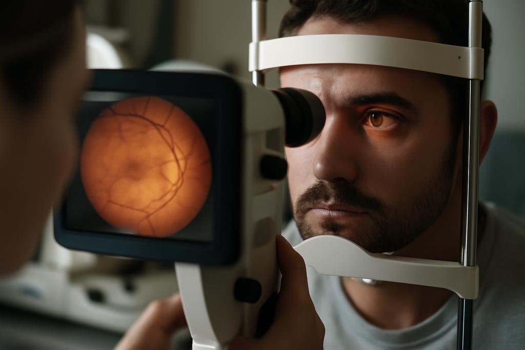
The human eye is a map of our health, a delicate mirror where tiny vessels tell big stories. Diabetic retinopathy, a complication that can creep up quietly for people with diabetes, damages those retinal blood vessels and threatens sight. Yet the gaze of the retina holds clues that can be read early enough to intervene. A team of researchers from Ahsanullah University of Science and Technology and Southeast University in Dhaka, Bangladesh, led by Shamim Rahim Refat and colleagues, set out to build a single, robust reader for that map. Their aim was not just to push accuracy on a single dataset, but to craft a system that could generalize across wildly different images – the kind of diversity you’d encounter in clinics around the world. In other words, they asked: can we teach a model to recognize the signs of diabetic retinopathy no matter where the photo comes from, what camera was used, or which country the patient calls home?
Their answer is a careful blend of data diversity, smart learning, and meaningful explanations. The researchers pull five publicly available retinal datasets into one hybrid collection that spans different imaging devices, populations, and severities. They also bring in techniques to balance the data so the model doesn’t simply memorize the majority class. And they pair this with a novel architectural idea that fuses the strengths of two established deep learning networks. The result is not just a higher score on a test bench; it’s a model designed to work in the real, messy world of clinical care while revealing the reasoning behind its predictions to doctors and patients alike.
A hybrid eye test: bringing five datasets under one roof
Medical researchers have long fought the twin demons of data bias and limited generalizability. A model trained on one collection of images may stumble when faced with a different camera, lighting, or patient population. The study tackles this by stitching together five public DR datasets: APTOS 2019, DDR, IDRiD, Messidor 2, and Retino. Each brings a unique mix of image quality, camera type, and DR severity distribution. The authors don’t pretend this fusion is trivial; they acknowledge how easy it is for a model to latch onto quirks of a single dataset rather than genuine disease signals.
Their hybrid dataset is meant to mimic the real world where clinics around the globe use different equipment and serve diverse patients. In their words, combining these sources “increases data diversity, reduces biases, and improves model performance across different clinical scenarios.” The payoff is a more resilient classifier that can handle the variation that often trips AI in medicine: variations in color balance, brightness, noise, and the telltale shapes of microaneurysms or hemorrhages that signal different DR stages. The team also applies a balancing technique called SMOTE to create synthetic examples for underrepresented DR stages, reducing the risk that the model will ignore rare but clinically important categories.
In addition to SMOTE, the authors use CLAHE, a smart contrast enhancement method that makes small features in fundus images – exactly the kind of details clinicians look for – more visible without amplifying noise. The result is a dataset that feels less like a collection of isolated images and more like a shared practice ground where features indicative of disease can be learned consistently across sources.
Two minds, one vision: fusing features from two networks
The centerpiece is a hybrid deep learning approach built to harvest the best of two different worlds. The researchers pair two established networks for feature extraction: one that excels at capturing fine-grained spatial details, and another that digs deep into abstract, high-level representations. The genius is not merely stacking them, but weaving their outputs together in a way that preserves both local texture and global context. The fused features are then refined by additional processing layers before a final decision is made about the DR severity class.
Think of it as two experts collaborating: one is a meticulous observer who notices tiny, local signs like microaneurysms or faint hemorrhages; the other is a strategist who understands the larger geometry of the retina and the progression of disease. By letting these two streams inform a shared classification, the model gains a richer, more balanced understanding of what distinguishes, say, no DR from mild DR, or moderate from severe. The result is a model that performs well across the five DR classes and can distinguish normal from abnormal images with higher confidence.
In their architecture, the fusion is not a blunt concatenation of features. The authors introduce a principled way to align and combine the feature maps so that the final representation remains compact and informative. The outcome is a dense, robust feature space that improves generalization across the heterogeneous hybrid dataset while keeping the computation manageable enough for practical use in clinics with limited hardware.
Making the decision legible: explainable AI for doctors
Clinical trust is won not just by accuracy but by the ability to understand why a model makes a particular call. The study places explainability at the forefront by applying five gradient-based explanation methods to highlight the regions of the retina that drive a prediction. Grad-CAM started the practice of visualizing where a CNN looks, but newer variants sharpen and localize the signal in sometimes surprising ways. Grad-CAM++, Layer-CAM, Score-CAM, and Faster Score-CAM each offer a different lens on the model’s attention, from broad maps to fine-grained heatmaps that home in on specific lesions.
In a medical context, these heatmaps aren’t mere curiosities; they act as a second opinion tool. If the AI flags a image as DR-positive or assigns a severity level, a clinician can inspect the heatmap to see whether microaneurysms, hemorrhages or exudates are driving the decision. The paper even discusses how forward-looking explanations can be paired with natural-language queries in a kind of AI-assisted diagnostic dialogue, which could help non-specialists interpret the results and discuss them with patients.
The upshot is not just a greener graph on a screen, but a tangible bridge between machine inference and human interpretation. In a field where subtlety matters and every patient’s retina tells a unique story, having interpretable, localized explanations can be as valuable as the prediction itself.
Numbers that tell a story about generalization
When the researchers tested single-model readers on each dataset, several architectures shone depending on the dataset. On APTOS 2019, for example, some networks reached very high accuracy, with some models hovering in the mid-90s. Messidor 2 and Retino told a similar story: the best architectures achieved impressive accuracy, yet the gains varied from dataset to dataset. This variance is exactly what the hybrid dataset seeks to address: a single model that performs consistently across diverse data sources.
On the big, blended challenge – the hybrid dataset – the landscape shifted. A two-network fusion model outperformed the individual networks across the board with an overall accuracy of 91.824%, precision of 92.612%, recall of 92.233%, and F1-score of 92.392%. The AUC under the ROC curve was a striking 98.749%, signaling that the model discriminates well across all DR levels. In short, the fusion approach not only held up but excelled in the presence of real-world data diversity, a crucial step toward reliable, wide-scale screening.
What’s especially interesting is the pattern across the broader family of networks. In several datasets, the older, more “classical” architectures that emphasize depth with careful local feature extraction delivered very strong performance, sometimes outperforming newer, more parameter-heavy models. The study’s message isn’t that bigger is always better; it’s that the right combination of strength matters. When you fuse complementary capabilities and train on a diverse canvas, you can build something that behaves well across the world rather than just in a lab.
Why this could reshape screening in the real world
Diabetic retinopathy is a candidate for large-scale screening precisely because the signs can be subtle and progressive. In many parts of the world, access to specialists who can read retinal images is limited. A robust, interpretable, generalizable model could empower non-specialists to screen patients, flag those who need urgent care, and track disease progression over time. The authors are explicit about a practical goal: a diagnostic tool that can function in resource-constrained settings, where cameras vary, lighting is imperfect, and clinicians need reliable guidance without sacrificing interpretability.
Beyond raw performance, the study’s emphasis on generalization and explainability has economic and ethical resonance. A model that performs consistently across populations reduces the risk of biased outcomes that can occur when a system is overfitted to a particular dataset. The explainability work helps clinicians trust the tool by showing them the retinal features that mattered most to the prediction. In a healthcare ecosystem that increasingly blends human and machine judgment, that level of transparency could be essential for adoption, regulatory acceptance, and patient comfort.
There’s also a pragmatic nod to the aging realities of clinical work. The authors discuss future directions that could improve both speed and robustness, including exploring vision transformer architectures that capture long-range context and using generative methods to balance underrepresented disease stages. While these ideas carry computational costs, they point toward a future where screening is both quicker and fairer across different patient groups and imaging environments.
From Bangladesh to the world: a human-centered achievement
Behind the numbers is a distinctly human story. The team’s insistence on harmonizing diverse data, their care in making the model’s decisions visible to clinicians, and their recognition of real-world constraints all reveal a project aimed at tangible impact, not just academic prestige. The study is anchored in universities with vibrant engineering and computer science programs in Dhaka, and it brings to light a collaborative spirit that crosses borders in the shared mission to protect sight. The lead author and co-authors, affiliated with Ahsanullah University of Science and Technology and Southeast University, demonstrate a regional strength that deserves wider attention as the field pushes toward scalable screening solutions worldwide.
The core idea is deceptively simple: if you want a system that helps people, it must understand people’s images in all their messy variety and explain its choices in human terms. The hybrid data strategy and the fusion-based architecture deliver that blend of robustness and clarity. In a health landscape where early detection can save sight and even lives, this work is an encouraging reminder that progress often comes from weaving together diverse sources of data and knowledge, then asking the machine to show its work in a way doctors can trust.
What this means for the future of eye health and AI in medicine
The study doesn’t pretend it has all the answers. It acknowledges limitations, notably the computational heft of larger models and the ongoing challenge of balancing datasets across all DR stages. It also looks ahead with humility: integrating vision transformers, leveraging GANs to expand the dataset with clinically plausible images, and bringing in multi-modal data such as patient history or genetics to improve accuracy and context. These are ambitious steps, but they map a credible path toward AI-assisted screening that is not only powerful but responsible.
Most of all, this work serves as a reminder that success in medical AI is not just about higher numbers. It’s about building tools that doctors can rely on, that patients can understand, and that can scale from a university lab to clinics in low-resource regions. The fusion of diverse data, the thoughtful pairing of feature extractors, and the emphasis on explainability together sketch a future where automated DR screening helps catch disease earlier, supports clinicians, and protects the gift of sight for millions around the world.


