


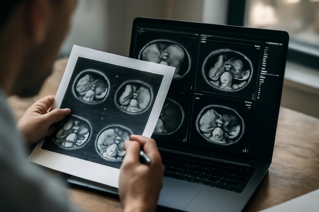
Radiology sits at the quiet edge of a medical emergency, a grayscale diary that chronicles what the body is doing at a moment in time. For the kidneys, those moments are often dense with subtle signs: a tiny cyst here, an exophytic nodule there, an attenuation value that can tilt a diagnosis one way or another. Historically, radiology reports have been written by humans who translate those pixels into sentences that tell clinicians what they should do next. Now, a team at the University of Florida is trying something equally ambitious and practical: teaching machines to read the slice by slice language of renal CT scans and to spit back human-readable notes that reflect clinically meaningful findings. The aim isn’t to replace doctors, but to augment their workflow with a second, highly trained observer that can standardize descriptions and reduce the time between scan and decision.
The study, led by Renjie Liang and Jie Xu from the University of Florida’s UF Health system and conducted with colleagues in the Department of Health Outcomes and Biomedical Informatics and the Department of Urology, represents a careful, clinically grounded foray into renal CT report generation. The researchers describe a two-stage pipeline in which a computer-vision model first extracts structured abnormality features from 2D CT slices, and a vision-language model then turns those features and the slice into a sentence that could sit in a radiology report. It’s a modular approach that mirrors how radiologists typically work: identify the clinically relevant attributes, then weave them into narrative that communicates risk, morphology, and potential next steps. What makes this work notable is not a dazzling new algorithm but a patient-centered insistence on clinical fidelity, interpretability, and real-world data constraints.
A Two-Stage Idea That Mirrors Real Work
The paper unfolds around a deceptively simple premise: kidneys speak in features, not just words. The first stage is a feature extractor that looks at each 2D slice and answers eight renal-specific questions: where is the lesion, how large is it, is it exophytic or not, what is its attenuation, does it enhance with contrast, is there a cyst, a mass, or a tumor? Some of these classifications are straightforward, others are trickier because real-world radiology reports are messy and incomplete. The researchers push back against the temptation to fill in missing data with defaults. If a feature isn’t described in the chart, they leave it unknown. That choice preserves realism and helps future work learn to handle real clinical ambiguity rather than pretend it doesn’t exist.
To train the tool, the team built a corpus from UF Health records, carefully pairing each sentence about a renal finding with the exact CT slice it describes and the feature labels that sentence implies. In total, they curated 130 annotated slices drawn from 97 patients. This isn’t a monster dataset by the standards of large public image collections, but it’s precisely the kind of domain-specific, clinicians-curated data that matters when you want clinical language to line up with image content. The lead authors and their collaborators annotated the triplets with a discipline-specific schema: position (left or right kidney), size in centimeters, exophytic status, attenuation (hypo-, iso-, hyperattenuating), enhancement, and whether a lesion is a cyst, a mass, or a tumor. When a feature was not described in the report, the label stayed Unknown. This preserves the reality that radiology notes are often incomplete, which in turn informs how well an automated system can perform in practice.
In the second stage, these structured features feed into a fine-tuned vision-language model designed to generate report-like sentences. The model’s prompt deliberately binds the language to the image, so the resulting sentence isn’t just fluent text but text that is anchored to a concrete slice and grounded in clinically meaningful attributes. The researchers emphasize alignment between the generated sentence and the ground-truth reporting, not just fluency or style. This is crucial: a sentence that sounds right but omits or distorts a finding could have real patient consequences. The system they built uses a modular architecture, which means future work can swap in more powerful readers or more precise language generators without remaking the entire pipeline.
Why This Matters for Patients and Radiology Teams
Kidney cancer and other renal abnormalities are on the rise globally, and CT imaging is a central tool for detection, characterization, and monitoring. As imaging volumes grow, radiologists face mounting pressure to produce accurate, consistent reports quickly. In that context, a system that can reliably extract salient features from images and translate them into clinically meaningful sentences could serve as a potent co-pilot. The University of Florida team is careful to frame their contribution as a feasibility study—the goal is to show that a modular, feature-informed approach can outperform a baseline that relies on chance or generic captioning, not to claim final clinical readiness. Still, the implications are meaningful: a workflow that could standardize certain phrases, ensure key attributes aren’t overlooked, and reduce the manual burden on radiologists would ripple through patient care by speeding decisions and improving consistency across readers and institutions.
Their results suggest that the feature extractor, when trained with appropriate task-specific losses and cross-validation, can detect several critical attributes with promising accuracy. Enhancement and cyst detection, in particular, show strong performance, and size estimation provides a rough but useful sense of lesion scale. The authors acknowledge the challenges of data imbalance and the fact that many features are Missing in real-world reports. That honesty matters because it signals where extra data collection, annotation effort, or algorithmic design might be needed before such a system can be deployed in a hospital’s radiology suite. The work also highlights how the two-stage design helps with interpretability: radiologists and researchers can inspect which features the model detected and how those features map to the final sentence. That kind of transparency matters when you’re building tools that clinicians will trust at the bedside.
What Surprised the Team and What Doesn’t Yet Hold Up
There are several notable takeaways about what the study achieved and where it still struggles. On the signal side, the system consistently outperformed a random baseline across all abnormality types. The model’s top performers include enhancement and cyst detection, with strong precision and F1 scores, which are clinically meaningful because enhancement can indicate vascular or lesion-specific differences and cysts are common but often require careful interpretation to distinguish benign from potentially worrying findings. Even in the attenuation task, where HU values can blur distinctions between hypo-, iso-, and hyperattenuation, the model showed meaningful gains over random guessing, hinting that the network can pick up contextual clues from surrounding tissue and reported language to refine its judgments.
But the study also offers a sober reminder of the limits imposed by small, imbalanced datasets. For a feature like exophytic status, the team faced extremes: only a single endophytic case in the annotated set, which inflates apparent performance due to memorization rather than generalization. They explicitly flag that issue, a rare but important moment of methodological candor in a field where hype around AI often outpaces data. The size estimation task likewise revealed sensitivity to how diverse the training samples are. In some folds, large lesions in the validation set challenged the model in ways not seen during training. These caveats aren’t roadblocks so much as guideposts pointing to what must improve before clinical deployment: broader data, more balanced representation, and perhaps new learning strategies that can generalize from few examples to rare but clinically critical scenarios.
Beyond the data caveats, the authors push back against overreliance on traditional natural language generation metrics. BLEU and ROUGE capture surface similarity but don’t guarantee clinical fidelity. The study’s own results show that while the generated sentences acheive reasonable lexical quality, there is still room to improve factual alignment with the image content in ways that physicians would explicitly rely on. That tension—between fluency and factual grounding—will shape how future renal radiology assistants are designed, tested, and validated. It’s a reminder that good medical language must do more than read like prose; it must hold up under the scrutiny of a clinician who may base a treatment plan on a single sentence in a report.
From 2D Slices to 3D Understanding and Beyond
One of the study’s most important limits is a focus on 2D slices. The authors deliberately ground their approach in the slice-level narrative that radiologists often reference in practice, but kidneys are three-dimensional objects, and tumors or cysts frequently span multiple slices. The authors envision a natural next step: extending the pipeline to full 3D CT volumes. That transition isn’t trivial. It would demand robust methods to fuse information across slices, maintain interpretability, and preserve the clinical grounding that makes the current approach valuable. Yet the payoff could be substantial: richer spatial context, better size and growth assessments, and the ability to generate more coherent, volume-aware reports that mirror how clinicians actually digest imaging data.
Beyond 3D, the authors point toward ways to strengthen evaluation and reliability. In medicine, human oversight remains essential, and as AI tools begin to generate sentences grounded in image features, the path to safer use likely runs through multimodal validation. That means not only refining generation metrics but also building frameworks for clinical evaluation: how often does the generated sentence reflect the actual findings, how does it influence clinical decisions, and how well does it integrate with electronic health records and existing reporting templates? The goal isn’t to manufacture more prose, but to align machine-generated language with the ethical and practical standards of medical care. This is unglamorous but essential work, and it’s where many of the hardest questions lie as the field moves forward.
Closing Reflections: Machines That Speak in a Doctor’s Language
What makes this study compelling isn’t a single breakthrough rumor about AI; it’s a careful construction of a workflow that respects the realities of clinical practice. The two-stage pipeline—first extracting grounded renal features from 2D slices, then generating sentence-level commentary with a vision-language model—feels like a practical blueprint for how to fold AI into radiology without displacing the clinician’s judgment. It’s an approach that foregrounds interpretability, data realism, and a modular design that can adapt as data quality improves and standards of care evolve.
At the heart of the UF effort is a simple, provocative idea: when a machine can describe a finding in precise, medically meaningful terms and attach those terms to a specific image location, you’ve built something closer to a collaborative tool than a black-box automaton. The study’s authors are not claiming a finished product; they are laying groundwork for a future in which renal reports could be more standardized, more transparent, and less burdened by repetitive drafting—without sacrificing clinical nuance. If such systems mature, they could help radiologists navigate rising volumes, give nephrologists and surgeons clearer summaries to anchor decisions, and even enable better patient-facing explanations of what a CT scan shows. In that sense, these kidneys are learning to talk in a language clinicians already understand—a promising step toward a more communicative, data-informed era of kidney care.
Lead researchers and institutions: The work is conducted by the University of Florida (UF) Health System, with contributions from the UF Department of Health Outcomes and Biomedical Informatics and the Department of Urology. The paper’s authors include Renjie Liang, Zhengkang Fan, Jinqian Pan, Chenkun Sun, Russell Terry, and Jie Xu, among others, reflecting a collaborative effort that spans imaging science, clinical practice, and health outcomes research. The study’s grounding in real-world UF Health data and its careful attention to clinical relevance serve as a reminder that the most impactful AI in medicine begins not in a laboratory alone but in a hospital, where data meet patients and clinicians meet needs.


