


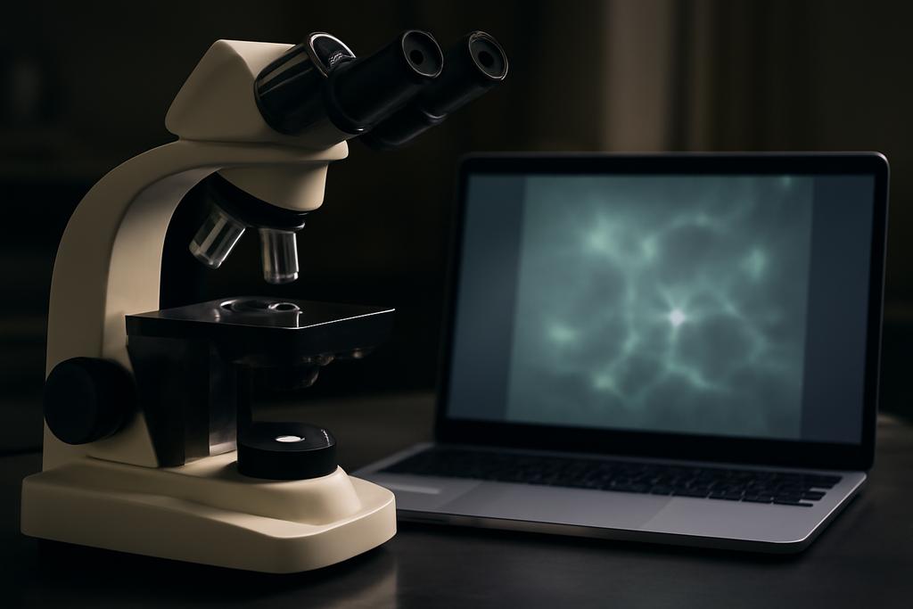
In biology labs around the world, the humble light microscope is a constant companion. It’s quick, cheap, and can reveal the tiny architecture of a cell, a neuron, or a budding organoid. But the very physics that makes it accessible also makes it hard to trust what you see. Widefield microscopes, the kind that sit on many benchtops, collect light from all depths of a sample. That means a lot of out-of-focus haze layers the image, softening edges and blurring the very structures scientists need to quantify. The expensive cure is optical: a confocal or other high-end setup that physically filters out the haze. The practical fix, increasingly, is computational: can a machine learn to produce a confocal-like image from a hazy widefield capture, without inventing new physics in the process?
That is the core question behind a study out of Human Technopole in Milan and Technische Universität Dresden in Germany, led by Anirban Ray and Florian Jug, with team members including Ashesh. They propose a method called HAZEMATCHING that sits at the crossroads of two long-standing goals in imaging: fidelity to the real data and realism in the sense that the results look like clean, plausible pictures of the same sample. The trick is not to pretend to “know” the exact, true 3D structure from a hazy image, but to learn a transport path from noisy, hazy observations to clearer, confocal-like predictions in a way that also acknowledges uncertainty. In other words, they’re not just dehazing; they’re dehazing with a conscious map of what could plausibly be true, and how confident the model is about each pixel.
The hazy truth behind widefield magic
To appreciate the challenge, picture a scene lit by uneven daylight through a dusty window. A photo taken through that window is softer and more ambiguous than a clean studio shot. In microscopy, the problem is amplified by the way light travels through tiny, complex structures. Widefield imaging captures both in-focus photons and a flood of out-of-focus photons from other depths, which act like a veil over the features scientists want to measure. Confocal microscopes handle this optically by using a pinhole to reject out-of-focus light. The result is sharper, crisper images with fewer blurring artifacts. But confocal systems are expensive, slower, and can be harsher on samples due to higher light exposure.
The interesting tension, then, is practical and philosophical. Do you push toward high pixel-level accuracy — a faithful reconstruction that minimizes numerical errors — or toward realism that makes the results feel like they belong to the same domain as the real, hazy training data? The former tends to produce outputs that look mathematically pristine but may miss subtle, biologically meaningful textures. The latter tends to generate striking, sample-like images that might tempt with convincing visuals but risk drifting away from the actual measurements. Blau and Michaeli’s perception-distortion trade-off, which many modern imaging papers invoke, crystallizes this dilemma: you can optimize for fidelity (distortion) or for perceptual realism, but not both equally well at the same time.
Balancing fidelity and realism in scientific imaging
HAZEMATCHING enters this debate not by picking a side, but by nudging the system to walk a middle path. It builds on a framework called conditional flow matching, a way to train continuous-time generative models that transport samples from a rough starting point toward a target distribution. The twist here is that the transport is guided by the hazy observation itself. In plain terms: the model learns how to turn random noise into a plausible, haze-free image, but it always uses the hazy input to steer that journey toward an answer that remains faithful to what the hazy image could logically reveal about the underlying sample.
The method operates with an input pair drawn from two imaging modalities: a hazy widefield image and its clearer counterpart captured with a more optical-precision setup. The research teams train the model with both synthetic data, where every variable is known, and real microscopic data, where the science matters most. A key design choice is to condition the transport not only on the hazy image but also on the per-sample content in the training data. This makes the dehazing story per-pixel and per-image, rather than a one-size-fits-all restoration.
Crucially, HAZEMATCHING does something most dehazing methods skip: it maintains an explicit pathway for uncertainty. Instead of returning a single “best guess,” it can generate multiple plausible dehazed outcomes, each corresponding to a different possible realization of the hidden, high-frequency details. In a field where noise and sample variation are the norm, this diversity matters. A single crisp restoration might feel clean but could obscure true biological variability. A distribution of plausible restorations, each grounded by the observed haziness, offers scientists a more honest set of possibilities to consider in downstream analyses.
HAZEMATCHING: guided flow as a new kind of microscope assistant
The core idea is both elegant and audacious. The model learns a time-dependent velocity field that, when integrated, transports a base random sample toward the non-hazy image distribution. But instead of learning the velocity field in a vacuum, it conditions the transport on the hazy observation xM0. During training, the model is fed triplets of data: a random noise sample x0, the hazy image xM0, and the clean target xM1. The training objective is to minimize the difference between the learned velocity and the true transport needed to morph x0 into xM1, all while staying guided by xM0. In simple terms, the neural network is learning the choreography for turning disorder into clarity, but it’s choreographing with the hazy image as a dance partner, not a solo dancer.
When it’s time to dehaze a new hazy image, the method starts from a fresh noise sample and uses the hazy image as the guide. The choreography unfolds via a small set of integration steps, essentially marching from noise toward a clean reconstruction by following the learned velocity field. Because the method is iterative, it can produce multiple plausible trajectories, yielding a collection of dehazed images rather than a single guess. The result is a family of predictions that share a coherent structure but differ in the fine details, a property that mirrors the natural variability of biological samples and the stochastic nature of photon-detection noise.
One of the paper’s appealing practical notes is that this approach does not rely on an explicit degradation operator to exist. In many diffusion-based or flow-based restoration schemes, you need to know exactly how the haziness was produced. In the wild, microscopy can have varied, sample-dependent hazing processes. HAZEMATCHING sidesteps that constraint, making it easier to apply to real-world data where the exact optics aren’t perfectly known or vary from experiment to experiment.
From a computational vantage point, HAZEMATCHING embraces a probabilistic mindset. Instead of chasing a single perfect image, it embraces a posterior view: after feeding in a hazy image, the method can generate a spectrum of credible, high-fidelity reconstructions. That spectrum is not just pretty graphics; it’s a quantified portrait of uncertainty that scientists can interrogate. The authors demonstrate that their method achieves a favorable balance on standard image-quality metrics that matter in microscopy, while also delivering well-calibrated uncertainty estimates. In other words, what you see is both sharper and more truthful about what isn’t certain.
Calibrated uncertainty and why it matters for science
A standout feature of HAZEMATCHING is its attention to uncertainty calibration. The team doesn’t stop at producing multiple plausible dehazed images; they also develop a way to align the model’s predicted uncertainty with actual errors observed on real data. They quantify, per pixel, how the predicted variability across samples correlates with the true errors. The result is a practical reliability map: if the model is uncertain about a region, a researcher can treat those pixels with caution, or design follow-up experiments to resolve the ambiguity.
That calibration is not cosmetic. In fields where decisions hinge on tiny, quantitative differences in structure — like the boundary of a cell nucleus, the integrity of a neurite, or the delicate architecture of an organoid — knowing when you might be wrong is as important as knowing what is right. It’s the difference between a botanist who trusts a single high-contrast photo and a scientist who knows which edges are well-supported by the data and which are still shadows in the data. The calibration work in HAZEMATCHING provides a bridge from raw pixel accuracy to scientific confidence, a bridge that could make dehazed images more trustworthy in downstream analyses such as segmentation, shape quantification, or disease-modeling studies.
Beyond the lab bench: why this could reshape scientific imaging
What makes HAZEMATCHING notable isn’t just that it clears hazy images. It’s that it reframes what counts as a good restoration in scientific work. In many imaging disciplines, there’s a natural impulse to calibrate toward the data you already have, rather than toward a glossy, “looks-like-a-clean-scientific-image” ideal. If a dehazing method can be tuned to respect the original measurements (fidelity) while offering multiple plausible clean views (realism) and a trustworthy map of uncertainty, it becomes a more honest partner for scientists who rely on those images to draw conclusions about biology and disease.
The use of a guided conditional flow framework also hints at a broader potential: cross-modality restoration without brittle degradation models, uncertainty-aware sampling that can inform experimental design, and iterative refinement that can be embedded into imaging workflows without forcing labs to upgrade every instrument at once. In a world where researchers are increasingly asked to extract maximum information from minimal photon exposure, a tool that can deliver crisp visuals while keeping uncertainty front and center could be a quiet revolution in how we collect, interpret, and trust images.
Limitations, caveats, and the road ahead
The authors are clear about the trade-offs. Iterative, sampling-based methods can be slower at inference than one-shot predictors. If a lab needs a single quick dehazing pass, there are faster baselines; if, however, a lab wants multiple high-quality candidates with calibrated uncertainty, HAZEMATCHING shines. The approach also relies on paired datasets for training, which may limit its immediate applicability to every imaging context. Yet the paper demonstrates robust performance across five datasets, including synthetic and real-world cases, suggesting a workable path to broader adoption as more paired data become available.
Another frontier is extending these ideas to other imaging modalities where hazy observations arise, such as electron microscopy, light-sheet imaging, or even non-biological scenes. The core insight — guided transport from noise to a target distribution under data-driven conditioning — is not tied to any one imaging modality. If researchers can assemble appropriate paired data or credible simulators, the same framework could help reveal hidden structure in many domains while keeping a clear line of sight to what is truly uncertain.
A new kind of clarity, born from collaboration between optics and algorithms
In the end, the study embodies a broader trend in science: the blending of physics-based instrumentation with data-driven inference to push the boundaries of what we can measure, without necessarily paying a premium in cost or sample toxicity. By asking the machine to respect both the hazy truth and the creative possibilities of what could be there, HAZEMATCHING invites researchers to see not just clearer pictures but also clearer reasons for the confidence (or lack thereof) behind each pixel.
As the authors note, the work came out of a collaboration between Europe’s Human Technopole and a nearby German university, with a global team of scientists contributing to the idea and its tests. The headline takeaway isn’t that we’ve invented a new filter or software trick; it’s that we’ve designed a way to navigate the space between what we measure and what we imagine, with a measured respect for uncertainty. For life sciences, that could translate into faster, cheaper, less invasive imaging workflows that still deliver trustworthy answers — a combination that feels as transformative as it is practical.
In practical terms, HAZEMATCHING offers a path to more accessible microscopy without sacrificing scientific rigor. It embodies a philosophy as old as science itself: seek clarity, but never pretend certainty where there is none. The creative twist is to let the uncertainty guide the search for clarity, rather than pretend it doesn’t exist. For researchers who routinely peered through hazy windows to glimpse the living world, this may be the most important kind of dehazing there is: a way to see more of what matters, while knowing what remains uncertain.
As the researchers behind the study acknowledge, the ultimate test is how these methods integrate into real-world workflows. The hope is that the combination of better visual realism, per-pixel uncertainty estimates, and a framework that does not rely on brittle degradation models will make such tools a friend to the bench — not a distraction from the science, but a dependable ally in the search for truth beneath the haze.


