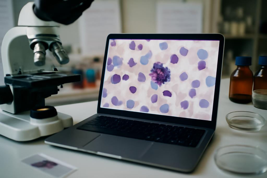
The study behind this piece tackles a quiet revolution happening at the crossroads of cancer biology and artificial intelligence. It asks a deceptively simple question with huge consequences: can machines reliably tell apart atypical mitoses from normal ones when the slides come from different labs, scanners, or even species? The answer isn’t a single yes or no, but a nuanced map of where today’s visual AI stands—and what we still need to build truly robust tools for pathology.
The work comes from a multinational team led by Sweta Banerjee at Flensburg University of Applied Sciences, with collaborators at the University of Veterinary Medicine Vienna and several other European institutions. They don’t just chase a high score on a single dataset. They assemble a rigorous benchmarking framework for atypical mitotic figure (AMF) classification, then test a spectrum of models across three held-out datasets that push the boundaries of domain shifts—from human breast cancer slides to canine tumors and real-world TCGA data. It’s a study about generalization as much as it is about accuracy, and it foregrounds a human-scale problem: counting and classifying mitoses is laborious for pathologists, and mislabeling or inconsistency can ripple into treatment decisions.
At their core, Banerjee and colleagues probe whether the latest, large-scale vision models—often trained on millions of slides and designed to generalize well—can actually handle the quirks of histopathology when data distribution changes. In medicine, the world doesn’t stay neatly the same from one lab to the next. Tissue preparation, staining, scanning hardware, and even species differences create subtle but meaningful shifts. The team’s answer is not a single verdict but a set of practical lessons about which strategies hold up in this messy real world.
What makes atypical mitosis meaningful
Amitotic figures (MFs) are the visual footprints of cell division. In normal tissues, they march through a well-ordered sequence—prometaphase, metaphase, anaphase, telophase—before two daughter cells emerge. Atypical mitotic figures, by contrast, display irregularities in chromosome alignment and segregation, a sign that the cellular machinery is misfiring. In cancer, such errors aren’t just curiosities; they’re prognostic breadcrumbs. A higher rate of AMFs has been linked—across some cancers and canine tumors—with more aggressive disease and poorer outcomes, making AMFs a potentially valuable biomarker for prognosis and treatment planning.
Yet identifying AMFs is no small feat. The features that separate an AMF from a normal MF can be subtle, and the labels themselves can be contested. The study builds on the idea that a reliable automated classifier must contend with two big realities: rarity and ambiguity. AMFs don’t appear as often as normal MFs on slides, and even experts disagree about borderline cases. In this work, the AMi-Br dataset, annotated by three pathologists (two board-certified), represents a consensus-driven effort to establish a robust training ground. Agreement across all three experts occurred in a little under 78 percent of cases, underscoring both the difficulty and the value of human guidance in training machines.
Beyond the human element, the biological stakes are clear. If we can quantify AMF prevalence across entire slides, we gain a potentially more objective read on tumor aggression. That could sharpen prognostic models and, in turn, influence how clinicians tailor therapies. The paper doesn’t claim to replace pathologists, but it points toward tools that can standardize measurements, reduce workload, and highlight areas where human review matters most. And in doing so, it frames a challenge that mirrors broader questions in medical AI: how do we build systems that understand biology but also survive the quirks of real-world data?
How the study tested AI across data walls
The authors don’t rely on a single problem or a single dataset. They introduce two new held-out AMF datasets—AtNorM-Br and AtNorM-MD—purpose-built to stress-test domain generalization. AtNorM-Br collects AMFs from the BRCA (breast cancer) arm of The Cancer Genome Atlas (TCGA), representing slides with mixed-quality images from multiple sources. AtNorM-MD expands the horizon to six domains, spanning human and canine tumors, drawn from the MIDOG++ training set. This multi-domain collection is the paper’s most provocative test, designed to reveal how well a model trained on one distribution can perform when faced with other tissues, species, and scanner histories.
On top of these datasets, AMi-Br remains the core training ground. It contains 3,720 MFs from human breast cancer, annotated by three experts and split into 832 atypical and 2,888 normal figures. The authors then subjected three families of models to the same binary task—AMF vs normal MF classification—across all three held-out datasets. The first family is end-to-end trained networks: EfficientNetV2, a Vision Transformer (ViT), and a Swin Transformer. The second family comprises foundation models—big, self-supervised ViT-based models trained on hundreds of thousands to millions of slides—with two probing strategies: linear probing (where the backbone stays frozen and a simple linear classifier is trained on top) and low-rank adaptation, LoRA, which fine-tunes only small, rail-thin adapters inside the model. A final trio of comparisons pairs five to eight different foundation models with these two adaptation strategies to cover a broad landscape of transfer learning options.
The evaluation is careful and transparent. They use five-fold cross-validation on AMi-Br, with slides (not patches) kept in separate splits to prevent data leakage. To address the class imbalance (far fewer AMFs than normal MFs), they optimize a balanced accuracy metric and employ weighted sampling during training. They report both balanced accuracy and AUROC (area under the receiver operating characteristic curve) to capture both outright accuracy and discrimination ability as decision thresholds shift. Across all experiments, the authors consistently stress domain shift as the central difficulty—how changes in staining intensity, scanner color profiles, or even species can erode performance.
What emerges from the data is a nuanced map of where different strategies shine. On the AMi-Br dataset, the LoRA-fine-tuned foundation models (especially Virchow2) reach an average balanced accuracy of about 0.8135, with AUROC just over 0.9. The Swin Transformer—an end-to-end baseline—earns a balanced accuracy around 0.805 and AUROC around 0.903, brushing the top of the field. But the Georgia-like story here is that the best-performing approach changes with the data. Across AtNorM-Br, the best result comes from an end-to-end ViT model, with balanced accuracy near 0.779 and AUROC near 0.871; the Swin Transformer is a close second. In AtNorM-MD, a more domain-diverse test, the end-to-end Swin again tops the table with a balanced accuracy around 0.772 and AUROC near 0.881, while Virchow, a foundation-model baseline tuned with LoRA, runs a strong second at about 0.770.
In plain terms, there is no single “best model” that wins across every dataset. The data shifts between labs, species, and tumor types tilt the playing field. The results also reveal that linear probing with foundation models—using a frozen backbone and a simple classifier on top—tends to lag behind LoRA-tuned or end-to-end methods, especially on the AMi-Br and AtNorM-Br datasets. Yet the foundation-models are not out of the game. They generalize to a meaningful degree and, in certain configurations, offer competitive performance with far less task-specific training. The subtle takeaway: bigger, pre-trained representations help, but you still need thoughtful adaptation to the target task and data environment.
What the results reveal about AI in medicine
One of the paper’s most striking conclusions is a reminder that generalization in medicine is not a solved problem by throwing more data at a model. Foundation models can offer robust, transferable features, but their advantage depends on how you adapt them to the target task. The LoRA approach—frozen backbones with small, trainable adapters—consistently improves performance across several models and datasets, sometimes dramatically. It’s a reminder that you can keep the heavy lifting in the big model but tailor the last mile to the job at hand with a fraction of the compute and data typically required for full fine-tuning.
Even so, the experiments also show a stubborn reality: domain shift remains a stubborn gatekeeper. The AtNorM-MD results reveal how performance can drop when the model faces a multi-domain, multi-species setup, even when trained on high-quality human breast cancer data. The authors point to a real-world implication: deploying an AMF classifier in a hospital or diagnostic lab is not a plug-and-play event. It requires careful curation of domain-relevant data, thoughtful calibration to local imaging conditions, and possibly a two-stage approach that couples automated detection with human oversight where the risk of misclassification is highest.
The authors also call out a subtle but important caveat: annotation biases can creep into the data. The AMi-Br dataset uses a three-expert vote to label mitoses, but even among trained pathologists, agreement is not universal. AtNorM-Br’s multi-expert labeling helps, but there remains a tension between perfectly reproducible ground truth and the messy, real world where slides vary by institution and scanner. The takeaway is not to pretend labels are perfect but to design benchmarks and models that acknowledge and survive this variability.
On the practical side, the study’s openness matters. The authors make their code and data publicly available, inviting the broader community to test, reproduce, and improve the benchmarks. This kind of transparency is essential if clinicians and researchers are to trust and adopt ML-assisted tools in the clinical workflow. The collaboration behind the work—across Flensburg, Vienna, Paris, Berlin, Ingolstadt, and beyond—also highlights how the biggest challenges in pathology often demand widescale, cross-institutional efforts rather than isolated lab-science wins.
What this means for the future of pathology AI
If you read the results as a single triumph for one model, you’d miss the bigger point. This work is a map of strengths and gaps, a candid inventory of how far today’s AI can travel when the road twists and turns from one dataset to another. The strongest takeaway is twofold. First, transfer-learning strategies that keep a large backbone fixed while learning small adapters (LoRA) can yield meaningful gains, particularly when data for the target domain are scarce. Second, end-to-end models—trained directly on the target task—often deliver the best performance on in-domain data and can remain competitive across domain shifts, provided you have enough annotated data to train them robustly.
What does that mean for real-world pathology? It signals that future AMF classifiers will likely be deployed as part of a two-stage system: a fast, robust detector that spots candidate mitotic figures, followed by a specialized, domain-aware classifier that discriminates AMFs from normal MFs with human oversight as needed. It also points to a broader design principle for medical AI: your system should reason about domain shifts explicitly, perhaps by training on multi-domain datasets, or by building models that can adapt quickly with small, task-specific updates. And it underscores the value of environmental diversity—datasets that span labs, scanners, and species—to train models that don’t fold under the pressure of real clinical variability.
Beyond the immediate task, the study is a striking case for how AI research in medicine can advance through deliberate benchmarking. The AMi-Br, AtNorM-Br, and AtNorM-MD datasets function as a public instrument—an invitation to test, compare, and push forward. The collaboration also shows how interdisciplinary teamwork matters: veterinary medicine, human oncology, computer science, and data science all come together to tackle a problem that lives at the edge of biology and technology. And it demonstrates the human element so often missing in techno-sudoku: behind every label, there are experts who spend years learning to read tissue with nuance, and their expertise remains essential even as machines learn to help, not replace, human judgment.
In sum, Banerjee and colleagues remind us that the future of pathology AI isn’t about a single silver bullet but about robust, thoughtful engineering that respects biology’s variability. The datasets illuminate how real-world data behave across species and sites; the models reveal a continuum between end-to-end learning and transfer learning that practitioners can tune to their needs. And perhaps most heartening, the work foregrounds open science as a path to steady improvement: a shared ground where researchers from Flensburg to Vienna and beyond co-create tools that could someday help doctors predict prognosis with more confidence and tailor therapies with greater nuance.
As one of the paper’s driving voices, Sweta Banerjee, and her team at Flensburg University of Applied Sciences show us a future where computational pathology learns not just the shapes of cells, but the shape of real-world uncertainty—where models grow more trustworthy as they learn to march through the messy, wonderful diversity of biology. It’s a future that feels less like a gadget and more like a collaborative instrument—one that could help clinicians read tumors with more clarity, across slides, labs, and species, and in doing so, sharpen the art and science of cancer care.
Lead researchers come from a remarkably collaborative roster: Sweta Banerjee and colleagues at Flensburg University of Applied Sciences (Germany) have spearheaded the AMi-Br benchmark, with crucial input from the University of Veterinary Medicine Vienna (Austria) among other partners. Their joint effort reflects a broader movement in which institutions such as Freie Universität Berlin, Technische Hochschule Ingolstadt, Medical University of Vienna, and Julius-Maximilians-Universität Würzburg contribute to a shared goal: teaching machines to understand cancer biology with care, humility, and scientific rigor.


