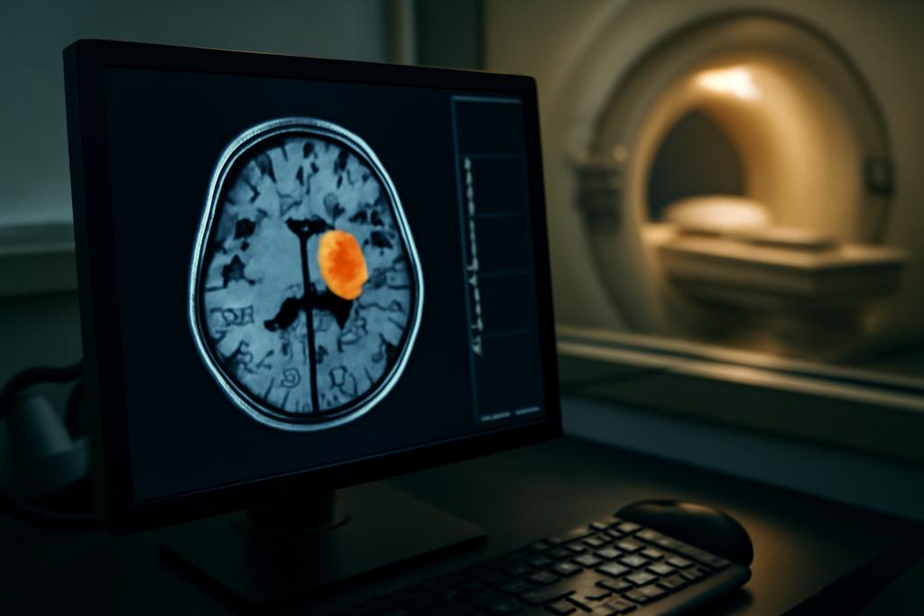
A Single MRI Image Learns to Segment Tumors On Its Own
In hospitals, the moment a patient walks in with a new MRI scanner or a fresh imaging protocol, the clock starts ticking for doctors and the artificial intelligence tools they lean on. Deep learning models trained on one set of machines often stumble when faced with a different brand, a different field strength, or a new protocol. It’s the phantom in the room: a model that shines in the lab but loses its glow when the environment shifts ever so slightly. The practical cost is real: slower workflows, more manual correction by clinicians, and in the worst cases, riskier decisions. This is not just a technical nuisance; it’s a barrier to turning smart imaging into reliable, per-patient help right at the bedside.
Enter a team from the Universitat de Barcelona and its Computer Vision Center collaborators, led by Smriti Joshi. Their goal is ambitious but elegantly simple: make a neural network adjust itself in real time, using only the single image it’s about to read. No need to collect a big, annotated target-domain dataset, no need to shuffle source data away from the hospital. The method aims to bridge the domain gap on the fly, so a patient’s own MRI becomes the tutor and the test image the classroom. If this works, it could be a practical game changer for medical imaging where patient-specific nuance matters as much as ever.
The paper, known as Single Image Test-Time Adaptation via Multi-View Co-Training, puts a spotlight on how to teach nets to cope with the unexpected by leaning on the very structure of medical volumes we routinely collect—3D, multi-view data that doctors already mentally sift through when judging a scan. The researchers ground their work in real clinical data: three publicly available breast MRI datasets, each with different vendors, scanners, and protocols. The study’s core claim is bold but specific: with only one test image, a model can adapt in one training epoch to achieve Dice scores that race close to the best fully supervised models trained with lots of data from the target domain. That’s the dream of test-time adaptation, realized for a highly practical, patient-level use case.
What makes this work stand out is not just the performance numbers, but the way it fuses several ideas into a coherent, lean recipe. The team calls their approach MuVi—short for Multi-View Co-Training. It lives in the space between self-learning from a single image and a principled use of the volumetric nature of MRI. Instead of asking for new labels or sprawling batches of target data, MuVi stitches together information from axial, coronal, and sagittal views of the same volume. It treats the target image like a puzzle where each view holds a piece that can guide the others, all while staying firmly in the patient’s own data—no leakage of the training set needed during inference.
Three-Dimensional Cues, One-Epoch Adaptation
Medical images aren’t flat pictures; they’re stacks of slices that form a landscape you can walk through. The authors exploit this by slicing the test volume into overlapping patches in three orthogonal directions: axial, coronal, and sagittal. Each patch carries a slightly different perspective—like looking at a sculpture from three viewpoints to understand its full shape. The method then asks the model to predict segmentation masks from each patch, while also encouraging the predictions from the alternate views to agree with each other. In practice, this means a patch, when transformed into its two other views, yields two additional “views” of the same underlying tissue. The training objective nudges the network toward a consistent view of the tumor across these perspectives.
Crucially, this co-training happens during test time and uses the target image itself. The goal is not to magically conjure labels or rely on pre-labeled data from the new domain; it’s to leverage the mutual information across views to sharpen the model’s understanding of the anatomy it’s looking at. A patch-level self-learning loss combines region-focused Dice loss with a cross-entropy term, but it’s the cross-view consistency that keeps the method grounded. Add a cosine similarity term between the feature embeddings of different views, and the network learns a representation that doesn’t snap back to one biased orientation when the data distribution shifts.
On top of this patch-based scaffolding, the researchers introduce an entropy-guided self-training mechanism. They compute uncertainty across the three views for each pixel and form a pseudo-label only when the confidence crosses a threshold. The thresholds aren’t one-size-fits-all; the highest-resolution view gets a stricter gate, while the other views have their own tuned thresholds. This prevents the system from bartering noisy predictions into the learning loop. The final objective, a weighted sum of the self-training loss, the cross-view consistency loss, and the embedding cosine loss, is designed to coax the network toward stability in the face of unseen shifts—without peeking at the source data during inference.
All of this is built on a solid, existing backbone: the 3D U-Net family popular in medical imaging, here adapted to work with a source network trained on a separate, larger breast MRI dataset. The authors deliberately keep BN statistics from the source intact during adaptation, adjusting only the scaling and shifting parameters (the gamma and beta terms). In other words, they let the model carry its learned sense of normality from its training domain, while teaching it to recalibrate to the new target domain using the patch views and the pseudo-labels produced on the fly. This isn’t a brute-force retraining; it’s a disciplined, per-image nudge in the right direction.
Why This Matters: Real-World Impact in the Clinic
Domain shift has always been the Achilles’ heel of medical AI. A model trained on data from one hospital may falter when deployed elsewhere, not due to a lack of intelligence but because the imaging pipeline changes—different scanners, different protocols, even different patient populations. The standard fix has been data collection and retraining, ideally with fresh annotations. In practice, that’s expensive, time-consuming, and often impractical in a hospital setting where decisions need to be made quickly and with patient-specific nuance. MuVi’s promise is to flip this dynamic: rather than waiting to accumulate a large pool of labeled data from every possible site, a clinician could feed the model a single patient image and watch the system adjust in real time.
From a clinical standpoint, the potential benefits are meaningful. First, the approach aligns with the realities of per-patient inference: you don’t need to bank on a multi-patient batch to make sense of a new scanner or protocol. Second, it leverages the volumetric nature of MRI. Rather than slicing the problem into 2D slices and risking a loss of 3D context, MuVi treats the volume as a whole tapestry of information that can be cross-referenced from multiple directions. Third, and perhaps surprisingly, the method demonstrates that you don’t necessarily need to trade off performance against uncertainty. By carefully balancing source-domain knowledge with target-domain adaptation through the beta/gamma modulation and view-based pseudo-labels, the team reports improvements that bring the target-domain performance close to a fully supervised upper bound—without needing target labels. In short, the model learns to see like a clinician who’s seen many scanners, but it does so with the patient’s own scan as the teacher.
The study’s authors—affiliates of the Universitat de Barcelona, the Computer Vision Center, and ICREA—tested their method on three publicly available breast MRI datasets that cover a spectrum of equipment and protocols. The Duke-Breast-Cancer-MRI data served as the source domain, while TCGA-BRCA and ISPY1 were used as target domains. Across these domains, MuVi outperformed several existing test-time adaptation methods and approached, or in some metrics neared, the supervised upper bound. The magnitude is not just a raw number; it represents a step toward reliable, low-friction deployment of AI in the clinic where data diversity is the rule, not the exception.
What Surprises and What Comes Next
There are a few striking takeaways that feel both technically interesting and practically provocative. One is the value of isotropic patches. The authors found that using patches with the same size across all three dimensions reduces directional biases that can creep in when you work with anisotropic, pancreas-to-brain style patches. In other words, treating the 3D volume with uniform blocks helps the model generalize better across different scanners and protocols. It’s a small change with outsized implications when you’re trying to bridge real-world domain shifts with limited target data.
A second surprise is the resilience of source-domain batch statistics. Several test-time adaptation methods lean on the target batch statistics to recalibrate normalization layers. But in single-image test time adaptation, you rarely have a reliable batch to draw statistics from. The MuVi approach keeps the source BN statistics intact and learns the per-image affine parameters (gamma, beta) to adapt. The result is more stable performance across diverse target domains, highlighting that sometimes the old data wisdom—trust the source distribution when you don’t have enough new data—still holds, provided you pair it with a principled adaptation strategy.
A third notable finding is the role of normalization when you push this kind of learning at the bedside. When the researchers experimented with Instance Normalization in place of Batch Normalization, the improvements were dramatic on the ISPY1 dataset, with DSC approaching the supervised upper bound and, in some metrics, even beating it. The HD and ASD numbers also improved meaningfully under this setting. This isn’t a universal win—normalization tricks often behave differently across datasets—but it underscores an important point: the right normalization choice can unlock robustness in ways that aren’t obvious until you test across real-world shifts.
All of this sits on solid experimental ground. The team evaluates across three datasets that differ in vendors, scanners, and planes, and they show consistent gains over several strong baselines, including normalization-based test-time methods and self-training approaches. The best-performing variant—MuVi with entropy-guided pseudo-labels and cross-view consistency—delivers a meaningful Dice-score gain over the baseline and narrows the gap to the supervised upper bound. It’s not a magic wand, but it’s a practical blueprint for making sophisticated segmentation tools more reliable when they meet the messy, variable world of real clinical data.
Looking ahead, the authors point to several exciting directions. Extending the approach to other volumetric imaging modalities—computed tomography, 3D ultrasound, or multi-sequence MRI—could magnify the benefits. There’s also a natural urge to explore hybrid strategies that blend per-image adaptation with selective access to lightweight, privacy-preserving target data when available. And as hospitals gradually deploy more edge-based AI systems, the prospect of fast, patient-specific adaptation without cloud dependencies becomes even more compelling.
As for the people behind the idea, this work foregrounds the collaboration between the Universitat de Barcelona and the Computer Vision Center, with Dr. Smriti Joshi taking a lead role among a team that includes Lidia Garrucho, Kaisar Kushibar, Dimitri Kessler, Oliver Diaz, and Karim Lekadir. Their collaboration spans mathematics, computer vision, and clinical insight, a reminder that the hardest challenges in medical AI often live at the intersection of disciplines rather than in a single field. The paper’s practical promise—quietly impressive performance gains from a per-image, on-the-fly adaptation technique—arrives at a time when clinicians and researchers alike are hungry for tools that can keep pace with the variability of real-world care.
In a world where data are precious, privacy is non-negotiable, and the single patient encounter is the most intimate data moment of all, MuVi offers a thoughtful path forward: let the patient’s own image teach the model how to see, moment by moment, without blasting the data across networks or waiting for large batches to accumulate. It’s a small, technically precise step, but it speaks to a larger aspiration: AI that moves as quickly and adaptively as a clinician who learns from every scan they read.
Note from the researchers: The methods described are designed to work within existing diagnostic pipelines and are released with code to support integration with nnUNet, a widely used medical-imaging framework, to help others reproduce and extend these ideas in real clinical contexts.


