


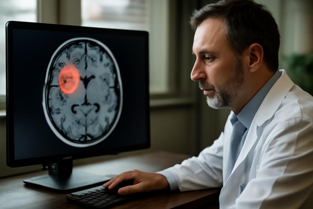
The race to perfect automated tumor segmentation in radiation therapy has taken a dramatic turn. For years, the challenge has been akin to finding a needle in a haystack – identifying cancerous tissue amidst the complex anatomy of a patient’s body. Manual delineation is painstaking, inconsistent, and time-consuming, demanding hours from already overworked radiation oncologists. Now, researchers at the University of Texas Southwestern Medical Center have introduced SAM2-Aug, a groundbreaking AI system that promises to revolutionize this crucial process.
The Power of Prior Knowledge
SAM2-Aug builds upon the impressive capabilities of the Segment Anything Model 2 (SAM2), a foundation model already renowned for its versatility in image segmentation. But the team didn’t stop there. They ingeniously integrated a powerful concept: prior knowledge. Think of it like this: instead of asking the AI to start from scratch with each scan, they gave it a head start. SAM2-Aug incorporates information from previous scans of the same patient, essentially providing the AI with a historical context to better understand the evolution of the tumor.
This is where the brilliance of SAM2-Aug truly shines. By leveraging this temporal dimension—previous images and their corresponding annotations—the AI doesn’t just see a snapshot; it perceives a dynamic picture of the tumor’s growth and movement. This contextual understanding dramatically improves its ability to pinpoint and delineate the tumor boundaries, even in cases where the contrast between the tumor and surrounding tissues is low or irregular.
Beyond Enhanced Accuracy
The impact of SAM2-Aug goes beyond simply improving the accuracy of tumor segmentation. The speed and efficiency gains are transformative. The ability to rapidly and reliably identify tumors using prior scans means less time spent on manual labor by oncologists, freeing them to focus on patient care and treatment planning. This is especially important in adaptive radiation therapy (ART), where treatment plans are adjusted daily based on the latest imaging, creating a high-stakes race against the clock.
Moreover, the study demonstrated that SAM2-Aug is remarkably robust and generalizable. It performs exceptionally well across various MRI sequences and different types of tumors, including liver, abdominal, and brain cancers. This means the system doesn’t need extensive retraining for each new scenario, making it a highly practical and versatile tool for a wide range of clinical applications.
The Methodology: A Symphony of AI Techniques
The researchers employed a clever combination of techniques to achieve these results. First, they enhanced SAM2’s input by providing it with a three-channel image: the current MRI scan, a prior MRI scan, and the corresponding tumor annotation from the prior scan. This approach provides both anatomical and contextual information to guide the segmentation process.
Second, they introduced a novel prompt augmentation strategy. Think of “prompts” as instructions given to the AI. The team didn’t simply provide static prompts; they incorporated random variations—expanding or contracting bounding boxes and using morphological operations on masks—to make the AI more resilient to imperfect inputs and less prone to overfitting.
Finally, they fine-tuned the parameters of the SAM2 model itself using a dataset of annotated medical images. This process allows the AI to learn the subtle nuances of medical imaging and adapt its segmentation capabilities specifically to the needs of ART.
The Results: A Clear Victory for AI
The results speak for themselves. SAM2-Aug significantly outperformed existing state-of-the-art segmentation methods across all tested datasets. It demonstrated superior accuracy and robustness, especially in accurately delineating tumor boundaries, even in complex and challenging scenarios. The improvements across various metrics—Dice score, Normalized Surface Dice, Hausdorff Distance, and Average Surface Distance—were consistently substantial, signifying a true leap forward in the field.
The study, led by You Zhang and a team at the University of Texas Southwestern Medical Center, is a testament to the power of integrating prior knowledge and leveraging the versatility of foundation models for medical image analysis. The researchers’ work opens up exciting new possibilities for improving the accuracy, efficiency, and accessibility of radiation therapy, ultimately benefiting countless cancer patients.
Looking Ahead: The Future is Promptable
While SAM2-Aug represents a significant advance, the researchers acknowledge areas for future improvement. They plan to explore ways to further refine the use of prompts, potentially reducing the number needed for accurate segmentation. They also aim to investigate the integration of 3D adapters within the SAM2 architecture to fully capitalize on the three-dimensional nature of medical images. This could lead to even more precise and efficient tumor segmentation.
The future of medical image analysis is clearly headed toward more sophisticated, promptable AI systems. The insights from this study are a significant step in this direction, showcasing the potential to transform cancer care through the power of AI-driven innovation. The code and trained models will be made publicly available at https://github.com/apple1986/SAM2-Aug.


