


Imagine trying to learn a new language, but all you have are fragments of conversations and a dictionary for a related tongue. That’s the challenge facing AI trying to diagnose a specific type of brain bleed, called aneurysmal subarachnoid hemorrhage (SAH). But what if that AI could ‘borrow’ knowledge from its experience with other brain injuries? That’s exactly what researchers at the University of Michigan are exploring, and their early results are promising. Led by Cristian Minoccheri, Matthew Hodgman, and Kayvan Najarian, the team is finding clever ways to teach AI to spot these subtle, often deadly bleeds, even when training data is scarce.
A Race Against Time in the Brain
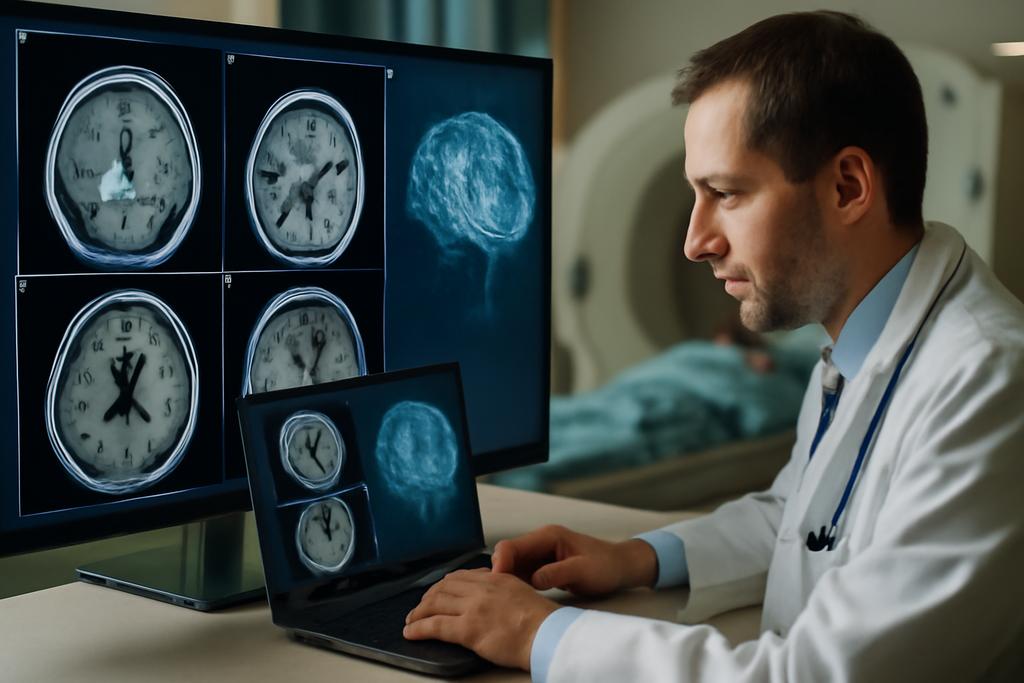
Aneurysmal SAH is a particularly nasty type of stroke where a blood vessel on the surface of the brain bursts. It’s a neurological emergency with a high mortality rate, exceeding 30%. The clock starts ticking the moment it happens; quick diagnosis and treatment are critical. Traditionally, doctors rely on CT scans to spot the telltale signs of blood in the subarachnoid space – the area between the brain and its surrounding membranes. But SAH can be tricky to identify, especially when the bleeds are small and scattered. It’s like trying to find faint constellations in a sky full of stars.
This is where AI comes in. Deep learning models, specifically those designed for image segmentation, have shown great potential in automatically detecting and outlining (segmenting) different types of brain bleeds. An AI that can quickly and accurately highlight these hemorrhages could be a game-changer, speeding up diagnosis and ensuring consistent care, especially in hospitals where specialists aren’t immediately available. The challenge? Training these AI models requires a mountain of labeled data – in this case, annotated CT scans showing exactly where the SAH is located. And high-quality SAH datasets are, unfortunately, hard to come by. Each scan needs to be reviewed and annotated manually by experts, a time-consuming and expensive process. This is where the concept of “transfer learning” becomes essential.
Borrowing Brainpower: Transfer Learning Explained
Transfer learning is a technique where a model trained on one task is repurposed for another related task. Think of it like this: if you’ve learned to drive a car, you’ll probably pick up driving a truck much faster than someone who’s never been behind the wheel. The AI equivalent involves pre-training a model on a large, readily available dataset – say, CT scans of patients with traumatic brain injuries (TBI), another condition involving brain bleeds. Then, this pre-trained model is “fine-tuned” on a smaller dataset of SAH scans. The AI leverages its existing knowledge of how blood appears on CT scans and adapts it to the specific characteristics of SAH. The beauty of transfer learning is that it allows us to get good performance even with limited SAH data. It’s like giving our AI a head start.
LoRA and DoRA: New Tools for Fine-Tuning
Now, here’s where it gets really interesting. The University of Michigan researchers didn’t just use standard transfer learning techniques; they experimented with some cutting-edge methods called LoRA (Low-Rank Adaptation) and DoRA (Weight-Decomposed Low-Rank Adaptation). These are “parameter-efficient” fine-tuning methods. Instead of retraining the entire AI model (which can be computationally expensive), LoRA and DoRA strategically tweak only a small subset of the model’s parameters. It’s like adjusting a few key knobs on a complex machine to get it working perfectly for a new task.
LoRA works by freezing the original model weights and adding small, trainable matrices (mathematical tables) to approximate the necessary adjustments. DoRA takes this a step further by decomposing the model’s weights into magnitude and direction components, allowing for more expressive and stable updates. Imagine you’re sculpting a statue. LoRA is like making small adjustments to the overall shape, while DoRA is like carefully refining both the size and angle of each individual detail.
The research team even developed a novel LoRA technique called CP-LoRA, designed to be even more parameter-efficient. They also introduced DoRA variants of existing LoRA methods, creating DoRA-C, convDoRA, and CP-DoRA. These new techniques represent a significant advancement in how AI models can be adapted for medical imaging tasks.
The Results: Impressive Gains in Accuracy
So, how did these methods perform? The researchers pre-trained a Unet architecture (a popular type of neural network for image segmentation) on CT scans from 124 TBI patients. Then, they fine-tuned this model on a dataset of just 30 SAH patients from the University of Michigan Health System. To ensure the results were robust, they used a technique called 3-fold cross-validation, where the data is split into three parts, and the model is trained and tested on different combinations of these parts.
The results were impressive. All the fine-tuning approaches, including standard methods, significantly outperformed a model with no fine-tuning at all. However, the LoRA-based methods consistently outperformed the standard Unet fine-tuning. In particular, a method called DoRA-C achieved the highest overall performance, demonstrating the power of these advanced adaptation techniques.
Interestingly, the researchers found that “over-parameterization” – using higher-rank adaptations that inject more parameters than strictly necessary – actually improved performance. This challenges the traditional assumption that low-rank adaptations are always the most efficient approach. It suggests that, in some cases, giving the model more flexibility can lead to better results, even if it means using more computational resources. It’s like giving an artist a wider range of colors to work with; they might not use them all, but having them available can lead to a richer, more nuanced creation.
Why This Matters: A Glimmer of Hope for Faster Diagnosis
This research demonstrates the potential of transfer learning and parameter-efficient fine-tuning methods to improve the accuracy and efficiency of SAH segmentation. By leveraging existing TBI datasets and employing techniques like LoRA and DoRA, AI models can be trained to detect these critical brain bleeds even when limited SAH data is available.
The implications are significant. Imagine a future where AI-powered tools can assist doctors in quickly and accurately diagnosing SAH, leading to faster treatment and improved outcomes for patients. This is especially important in smaller hospitals or rural areas where access to specialized neuroradiologists may be limited. An AI could act as a “first responder,” flagging suspicious scans for further review by a human expert. While the study acknowledges limitations, like the small dataset size and single-institution data, it represents a crucial step forward in the quest for automated SAH diagnosis.
The University of Michigan team’s work highlights the power of creative problem-solving in AI research. By thinking outside the box and borrowing knowledge from related domains, they’re paving the way for a future where AI can play a vital role in saving lives and improving healthcare for all.


