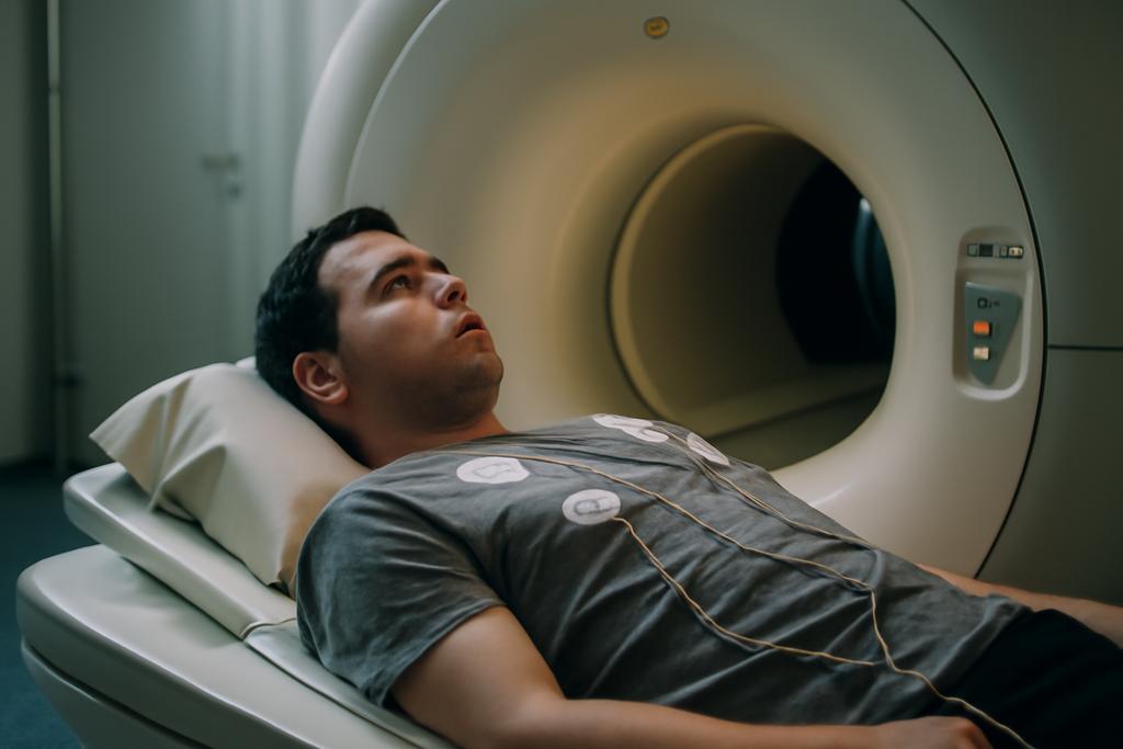
A Race Against Time: The CMRxMotion Challenge
Cardiac magnetic resonance (CMR) imaging is the gold standard for evaluating heart health, offering detailed views of the heart’s structure and function. But like a seasoned musician needing perfect silence to hit a high note, CMR scans require patients to hold their breath precisely. Any involuntary movement—the slightest breath—introduces artifacts into the images, degrading their quality and potentially skewing diagnostic results. This is a significant challenge, especially with patients who struggle with breath-holding, such as those with heart failure or children. The problem: how do we make sure that AI diagnostic tools aren’t tripped up by these breathing hiccups?
The Stakes Are High: The Need for Robust CMR Analysis
The increasing use of artificial intelligence (AI) in medical image analysis promises to revolutionize healthcare, enabling faster and more accurate diagnoses. In CMR imaging, AI algorithms can automatically segment the heart’s structures—such as the left and right ventricles and the myocardium—extracting crucial clinical biomarkers like ejection fraction (the percentage of blood pumped out of the heart with each beat) and ventricular volumes. These measurements guide treatment decisions, making the accuracy of the algorithms paramount. However, if these AI tools are trained primarily on pristine images, their performance can dramatically plummet when presented with the kinds of real-world imperfections that routinely arise in clinical practice. A CMR scan ruined by respiratory motion might need to be repeated, adding extra time and discomfort for the patient.
A Novel Challenge: Engineering Real-World Imperfections
To push the limits of AI’s ability to accurately diagnose from imperfect images, researchers at Fudan University, Imperial College London, and several other institutions organized the CMRxMotion challenge. Instead of relying on existing, often messy datasets with confounding factors like varying scanner models and patient conditions, they decided to carefully engineer the problem. They recruited 40 healthy volunteers and, using the same 3T MRI scanner (Siemens MAGNETOM Vida), instructed them to perform CMR scans under varying breathing conditions: perfectly holding their breath, halving the breath-hold period, breathing freely, and breathing intensively. This created a controlled dataset of 320 CMR cine series with a spectrum of motion artifacts, ranging from none to severe. Lead researchers on the project included Chengyan Wang, Wenjia Bai, and Shuo Wang.
Two Tasks, One Goal: Evaluating AI’s Resilience
The challenge consisted of two parts, both designed to test the limits of AI: The first task was automated image quality assessment (IQA): could algorithms reliably determine the severity of motion artifacts in the images? The second task was robust cardiac segmentation (RCS): could algorithms accurately segment the heart’s structures, even with imperfect images? In total, 22 teams from around the world submitted algorithms.
The Results: A Mixed Bag of Successes and Challenges
The results were interesting. In the IQA task, the best-performing algorithms demonstrated good agreement with the assessments of expert radiologists, suggesting that AI could be a useful tool for preliminary image quality control. However, a key limitation was the difficulty in accurately classifying the severity of motion in severely degraded images. While AI excelled at identifying high-quality scans, it lagged behind when it came to pinpointing the exact degree of motion artifacts.
The RCS task yielded surprisingly high accuracy in images with minimal motion artifacts. The top-performing algorithms were often on par with human experts in segmenting the left ventricle and myocardium (the heart muscle). However, as the severity of motion increased, so did the error rate. The error in crucial clinical biomarkers derived from the segmentation—like left ventricular ejection fraction—grew exponentially with increasing artifact severity. This highlights a crucial point: even small errors in segmentation under imperfect conditions can lead to inaccurate clinical interpretations with potentially serious consequences.
Lessons Learned: The Path Forward
The CMRxMotion challenge offers several valuable lessons for the field. First, it highlights the urgent need to develop AI algorithms that are truly resilient to the kind of real-world imperfections that are encountered in routine clinical practice. Second, it suggests that while AI has the potential to aid in preliminary image quality assessment, it still struggles to assess fine-grained differences in image quality. Third, it emphasizes the need to balance AI model accuracy with computational efficiency to facilitate clinical implementation.
The future of AI-powered CMR analysis hinges on addressing these issues. This includes developing more sophisticated architectures, focusing on computational efficiency, and collecting large-scale, multi-center datasets with a wider range of real-world scenarios. By acknowledging the limitations of current AI and the importance of robustness, the research community can work toward truly reliable tools for improving healthcare.


