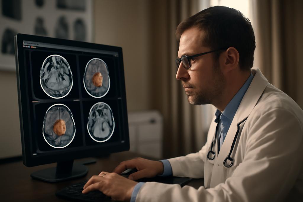
Imagine a world where medical image analysis is not a laborious, specialized task, but a swift, reliable process. That’s the promise of UniSegDiff, a groundbreaking new model developed by researchers at Dalian University of Technology, led by Lihe Zhang. This advanced AI system utilizes a ‘diffusion model’—a type of artificial intelligence that learns by adding and removing noise from images—to achieve unprecedented accuracy in detecting and segmenting lesions across a wide variety of organs and imaging modalities.
The Challenge of Unified Lesion Segmentation
Current methods for detecting tumors or other lesions in medical images are often highly specialized. A model trained to identify lung tumors on CT scans might not work effectively on identifying polyps in colonoscopy images. This specialization creates bottlenecks and inconsistencies in medical image analysis. What’s needed is a unified system—a kind of medical image Swiss Army knife—that can adapt effortlessly across different organs, image types, and data sets.
The problem isn’t just about the diversity of images. Think of the challenge like this: finding a specific type of sea creature in the ocean is hard enough when you’re looking at just one type of coral reef. But if you had to hunt the same creature across all the diverse environments of the world’s oceans—from the Arctic to the tropical, the shallows to the deepest trenches—that’s an exponentially harder task.
This is precisely the challenge that UniSegDiff tackles. It aims to create a single model capable of consistently identifying various lesions across a diverse range of medical imaging data.
Diffusion Models: A New Approach to Medical Image Analysis
UniSegDiff employs a relatively new type of AI called a diffusion probabilistic model (DPM). Think of DPMs as a clever way to train an AI to ‘see’ by gradually adding noise to an image and then learning how to reverse that process to clean it back up. It’s as if you were to slowly smudge a painting, and then learn to reconstruct the original by gradually un-smudging it, layer by layer. The randomness inherent in the process helps the model avoid overfitting to the nuances of a specific dataset, allowing it to generalize better to unseen data.
However, previous attempts to use DPMs for lesion segmentation faced challenges. Existing models tended to focus their attention disproportionately on certain parts of the training process (like the noise-reduction phase), leaving other crucial steps underserved. This led to longer training times and less-than-optimal results.
UniSegDiff: A Staged Approach
The researchers overcame this limitation by cleverly dividing the training process into three distinct stages, each with a specific focus:
- Rapid Segmentation Stage: This initial phase focuses on quickly generating an initial segmentation map. Think of this as a rough sketch.
- Probabilistic Modeling Stage: Here, the model refines its understanding of the lesion’s characteristics, taking into account both the image and the noise during the denoising process.
- Denoising Refinement Stage: This final stage focuses on finely tuning the edges and details of the segmentation, similar to adding those finishing touches to a painting.
This staged approach ensures that the model pays attention to all aspects of the training process, resulting in a more comprehensive and accurate understanding of the images. It’s like having a team of artists, each with a specialized role, working together to create a masterpiece.
Pre-training and Uncertainty Fusion: Enhancing Robustness
To further enhance its capabilities, UniSegDiff also employs pre-training. Before it even begins training on lesion images, the model is trained on a large dataset of images from various sources. This ensures that the model has a strong foundational understanding of medical imaging before it starts tackling the complexities of lesion segmentation.
Finally, UniSegDiff uses a process called uncertainty fusion. The model generates multiple segmentation results, and then intelligently combines them to arrive at the most accurate final segmentation. This approach greatly increases robustness and avoids the potential pitfall of relying on a single, potentially flawed, prediction.
Results and Implications
The results are astonishing. UniSegDiff significantly outperformed all existing state-of-the-art methods across six different organs and image modalities. The fact that it performs so well across diverse datasets represents a significant leap forward in the field. The impact of this could be enormous. It could lead to faster, more consistent diagnoses, saving valuable time for doctors and improving patient care.
Looking Ahead
The researchers are already working on expanding the model’s capabilities to include more organs and types of medical images. They are also exploring ways to extend UniSegDiff to work with 3D images, further enhancing its potential applications. The future looks bright for this powerful new tool in the fight against disease.


