The Murky World of Medical Images
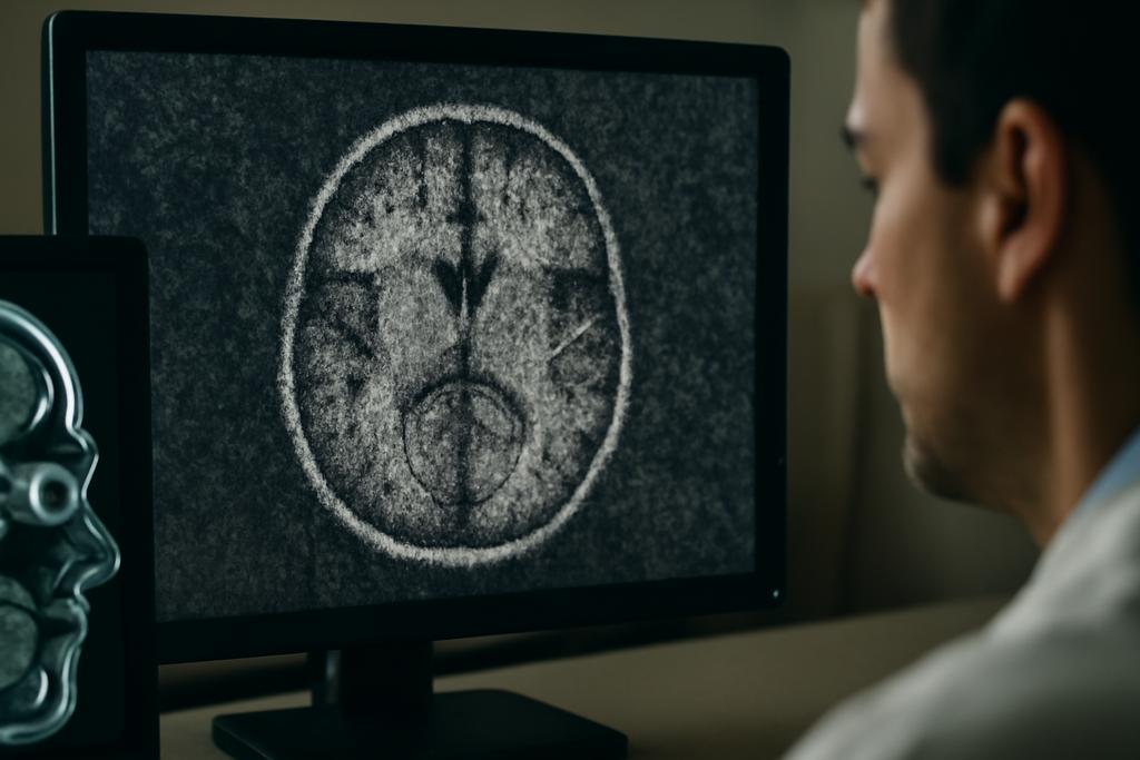
Medical images—the X-rays, CT scans, MRIs that illuminate the inner workings of our bodies—are rarely perfect. Noise, a kind of static inherent in the imaging process itself, muddies the picture, making it harder for doctors to make accurate diagnoses. This noise isn’t just a minor annoyance; it can be a significant obstacle, potentially leading to missed diagnoses and compromised treatments. It’s like trying to read a crucial document with smudges obscuring key words; the message is there, but hard to decipher.
Cleaning Up the Static: A New AI Approach
Researchers at Aston University, led by Jitindra Fartiyal and Sergei K. Turitsyn, have developed a new AI-powered method to dramatically improve the clarity of these images. Their technique, called Dual-Path Learning (DPL), tackles the problem head-on by using a sophisticated neural network approach. Instead of simply trying to filter out noise using blanket methods, DPL uses two separate, parallel pathways to analyze the image.
One pathway focuses on pinpointing the noise itself. Imagine a detective scrutinizing a crime scene for clues. This pathway carefully analyzes the image to identify and characterise the nature of the noise: where it’s most prominent, its texture, and intensity. The second pathway simultaneously focuses on the underlying structure and context within the image, similar to an artist carefully observing the subject, attempting to separate important details from distractions.
These two pathways work together – much like two detectives comparing their observations – providing the algorithm with rich, complementary information about the image. By fusing these sources of knowledge, the DPL algorithm constructs an image significantly clearer than methods that try to accomplish the same task through single-pathways.
Beyond Single Images: A Universal Approach
Existing AI denoising methods typically focus on a single type of image and one specific type of noise, like trying to fix a particular brand of TV with a universal remote. The groundbreaking aspect of DPL is its remarkable versatility. Aston University’s researchers tested their algorithm on a diverse range of medical images—CT scans, MRIs, optical coherence tomography (OCT) scans, and fundus images—with various kinds of noise contaminating the data. This approach is like designing a pair of glasses that can correct several types of vision problems at once, without any adjustment needed.
Measuring Success: Beyond the Numbers
The researchers used standard metrics to evaluate the performance of DPL, such as PSNR (Peak Signal-to-Noise Ratio) and SSIM (Structural Similarity Index). These metrics quantify how much the algorithm improves the image quality, comparing the processed image to a clean version. DPL consistently outperformed not only traditional image-processing techniques, but also other cutting-edge AI-based methods, producing substantially clearer images. This is particularly important in cases where even minor noise can have huge consequences.
However, the story is much more nuanced than just those numbers. Visual inspections showed that DPL not only produced images with better numerical scores, but also images that were more visually appealing, retaining fine details that might otherwise be lost. In other words, DPL did more than just improve the numbers; it improved what actually matters for diagnosing patients – the overall quality of the image.
The Real-World Impact: A Clearer Picture of Health
The implications of DPL are vast. In clinical settings, clearer images could mean earlier and more accurate diagnoses, better treatment plans, and ultimately, improved patient outcomes. This would be particularly helpful in situations where a low dose of radiation is preferred, leading to lower-quality images. DPL could provide a solution to this problem. The improved image quality may also improve the performance of AI algorithms used in medical image analysis, providing even more accurate diagnostics.
Looking Ahead: Expanding Horizons
While DPL shows immense promise, the researchers acknowledge some limitations. The current study used relatively small datasets for some imaging modalities. Future research will involve expanding the datasets and refining the algorithm to further improve its performance on less common image types. The researchers are also eager to test their method in real-world clinical settings to assess its full impact on diagnosis and treatment.
The development of DPL represents a significant leap forward in medical image analysis. This powerful AI technique has the potential to transform the healthcare landscape by providing doctors with a clearer and more accurate picture of their patients’ health, all thanks to a clever method for removing noise in images.


