


Medical imaging is a cornerstone of modern healthcare, but interpreting images can be slow and subjective. For abdominal scans, the gold standard is often a laborious, manual process, prone to errors in consistency. Now, a new benchmark study from Brigham and Women’s Hospital, led by Deepa Krishnaswamy and Cosmin Ciausu, has evaluated the most promising AI methods to automate this task, revealing both impressive progress and subtle but crucial limitations.
The Challenge of Abdominal MRI Segmentation
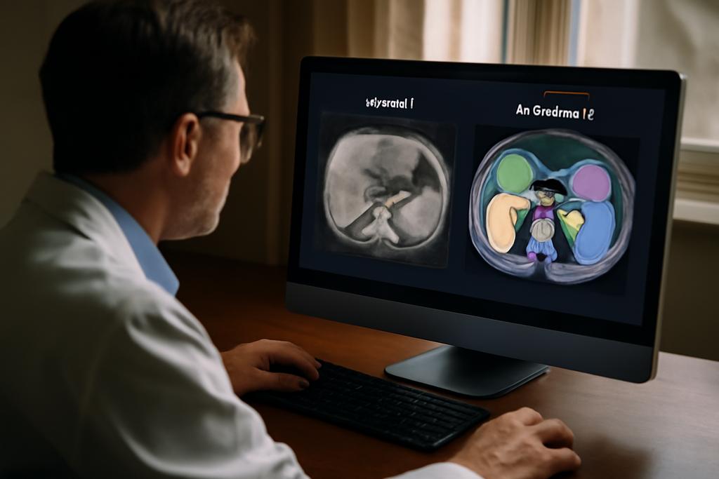
The human body is remarkably complex, and creating a perfect 3D map of its interior from images is a far more difficult challenge than most people realize. Think of it like this: you’re given a blurry photograph of a city skyline at dusk, and you have to identify every building, determine its size, and map its location perfectly. That’s essentially what scientists are tackling with MRI scans of the abdomen. Compared to CT scans, MRIs offer a far more nuanced view of tissues, but their inherent signal variability makes automated analysis far more challenging.
This variability is due to the multitude of parameters involved in acquiring MRIs. Every image is unique, affected by many variables during the scan. The signal isn’t as standardized as CT scans, making the creation of AI models that can consistently interpret these differences notoriously difficult. This has traditionally hindered the development of reliable, automated systems for segmenting (identifying and separating) abdominal organs.
Deep Learning to the Rescue (Mostly)
Recent advances in deep learning — a branch of AI that uses neural networks to learn patterns in massive datasets — have given us the ability to train algorithms that can perform complex image analysis. Three state-of-the-art models were evaluated in this study: MRSegmentator, MRISegmentator-Abdomen, and TotalSegmentator MRI. These models are incredibly sophisticated, capable of identifying and isolating dozens of different anatomical regions, from the liver and spleen to the kidneys and the adrenal glands. However, training these algorithms requires enormous amounts of accurately labeled data — essentially, having a human expert meticulously outline each organ in thousands of MRI scans. This is incredibly time-consuming and expensive.
The Synthetic Solution: ABDSynth
Because creating these annotated datasets is such a huge undertaking, scientists have begun to explore a clever alternative: using synthetic data. This study introduced a new method, called ABDSynth, which leverages a technique known as SynthSeg. This method uses existing CT scans (which are more readily available and already well-annotated) to create synthetic MRI scans. The AI model is then trained on these artificial scans, which effectively allows it to learn the patterns needed to interpret the far more challenging real MRI images without requiring any actual human annotation of MRI scans.
The implications here are profound. While traditional models might require thousands of meticulously labelled MRI scans and human expertise to refine labels, ABDSynth offers a shortcut. It’s like having a digital artist create a near-perfect replica of a famous painting, but for organs. This could significantly reduce the time and cost associated with developing these vital tools.
The Verdict: Strengths, Weaknesses, and the Future
The researchers evaluated these models on three large, publicly available datasets, ensuring their findings generalize beyond specific hospitals or scan types. Overall, MRSegmentator achieved the best performance, consistently producing accurate and reliable segmentations. However, even the top-performing model struggled with smaller organs and highly variable regions, highlighting the challenges inherent in the task.
ABDSynth, the synthetic model, performed surprisingly well, especially considering it was trained entirely on data from a different imaging modality. While slightly less accurate than the best real-data models, its reduced reliance on manual annotation makes it a very promising option, especially for applications with limited resources. This method highlights the potential of synthetic data to bridge the gap between the need for vast, high-quality datasets and the reality of medical data availability.
The study also highlighted an important limitation: inconsistencies in how organs are defined and labeled. This discrepancy illustrates a broader issue in medical image analysis — the need for standardization across datasets. Without such standards, even the most accurate AI algorithms can struggle to generalize across different contexts.
The Larger Picture
This research is more than just a technical achievement; it’s a significant step forward in making advanced medical imaging accessible and affordable. By improving the accuracy and efficiency of organ segmentation, we can accelerate the diagnosis and treatment of a wide range of abdominal conditions. The ongoing quest for more efficient and robust algorithms, fueled by both real and synthetic data, will undoubtedly lead to better patient care in the years to come.
The work underscores that even the most advanced AI algorithms are not a silver bullet. They require careful evaluation, attention to data quality and consistency, and recognition of their inherent strengths and limitations. Nonetheless, the findings offer a compelling vision of a future where AI-powered image analysis seamlessly integrates into clinical practice, improving both the quality and efficiency of healthcare.


