


Foundations Enter Pathology, Changing the Data Game
Highlights: Big neural networks trained on unlabeled data can unlock delicate pathology tasks with far less labeled data.
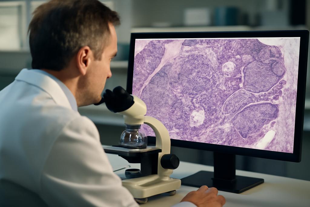
When you look at a whole slide image from a tumor tissue, you’re staring at a billion little pixels that tell a story—one that pathologists have learned to read through years of training. The new study by a multinational team led by Technische Hochschule Ingolstadt and collaborators across Germany and Austria asks a simple, ambitious question: can the giant, self supervised foundation models trained on oceans of unlabeled images be repurposed to identify mitotic figures—tiny, crucial markers of tumor aggression—without drowning in the label-crafting process? The answer, in short, is yes, but with some elegant caveats. The authors—headed by Jonas Ammeling and a team that includes Marc Aubreville and Katharina Breininger—show how these models, once adapted with smart, data-efficient techniques, can reach surprisingly strong performance even when labeled data are scarce.
In this collaboration, six pathology-focused foundation models were pitted against traditional baselines on two public datasets. The project isn’t just about chasing top numbers; it’s about whether you can build robust, general-purpose vision systems for pathology that don’t crumble when you move them from one lab to another. That’s where the science becomes personal for clinicians and patients alike: a model that needs a hundred times fewer labeled examples to learn a task could accelerate translation from research to real-world clinics, and potentially reduce the wait for faster, more accurate diagnoses.
Behind the study is a constellation of institutions—the Technische Hochschule Ingolstadt’s AImotion group, MIRA Vision Microscopy, the University of Veterinary Medicine Vienna, Julius-Maximilians-Universität Würzburg, Friedrich-Alexander-Universität Erlangen-Nürnberg, and Flensburg University of Applied Sciences—embodied in a cross-disciplinary riff between computer science, veterinary pathology, and biomedical engineering. The lead author is Jonas Ammeling, with a diverse set of co-authors including Emely Rosbach, Ludwig Lausser, Christof A. Bertram, Katharina Breininger, and Marc Aubreville. The ensemble reminds us that the future of AI in medicine is being built not in one lab, but in a network of labs, clinics, and companies sharing a common goal: making high-stakes histology safer and more scalable for real patients.
Why data scarcity has haunted pathology—and how SSL helps
Highlights: Self-supervised learning unlocks learning from unlabeled slides, leveling the playing field when experts are scarce.
Pathology has always walked a tightrope between data abundance and data scarcity. High-resolution whole slide images are plentiful in the wild, but high-quality labels are scarce, time-consuming, and costly. Pathologists’ fatigue, the variability in staining across centers, and the sheer size of WSIs make curated labeling a heavy lift. The paper frames this as a practical bottleneck: even as AI models get smarter, their best performance still hinges on how much carefully annotated data you can afford to produce.
That’s where self-supervised learning—the idea of teaching a model to understand structure in data without explicit labels—becomes a lifeboat. By training on billions of unlabeled tiles, modern foundation models learn rich, transferable representations. Think of SSL as giving the model a sense of “grammar” for images: it learns what makes a patch look like a tissue, what patterns belong to cells, and what tissues tend to look like under different magnifications. The downstream task—mitotic figure classification—then becomes a matter of teaching a light, task-specific reader to use those deep representations, rather than starting from scratch with every label.
The paper’s precision is in how it tests two pathways to leverage those representations: linear probing, where a tiny linear classifier sits on top of frozen features, and LoRA, a parameter-efficient fine-tuning method that nudges parts of the model via low-rank updates. The contrast isn’t merely academic. It translates into real differences in how quickly a model can adapt when labeling budgets are tight and when you’re migrating a model from one lab to another with different scanners and staining. The authors also deliberately compare these foundation-model approaches to traditional, end-to-end trained networks, reminding us that the best tool often depends on the data regime you’re operating in.
In short, this section isn’t a tribute to bigness for its own sake. It’s about learning how to bend the scale of pretraining and adaptation to make a clinically useful tool that’s robust where it matters: in the wild, across centers and even across species in some tasks. The human experts behind this work aren’t just pushing numbers; they’re mapping a practical path toward dependable AI-aided pathology that can weather the real-world messiness of a multi-center study in the wild. The foundation models aren’t magic; the innovation is in how you tune, adapt, and validate them against tasks that matter to patients and pathologists alike.
Two datasets, six models, one question: how well do we scale?
Highlights: The benchmark uses two diverse datasets and shows how model scale and adaptation interact with data availability.
The authors anchor their analysis in two well-chosen public datasets that together span a spectrum of real-world challenges. CCMCT is a large, single-domain resource focused on canine cutaneous mast cell tumors, with tens of thousands of mitotic figure annotations. It’s a dense, single-tayload scenario—a good stage for data-scarce to data-plentiful comparisons from a controlled biological context. MIDOG 2022, by contrast, is a multi-domain, multi-tumor, multi-laboratory corpus that includes species and scanner variety. It’s the kind of dataset that exposes a model to distribution shifts it will encounter in actual clinics: different slide preparations, different equipment, and different biological manifestations across tumor types.
From there, Ammeling and colleagues evaluate six pathology foundation models—Phikon, UNI, Virchow, Virchow2, H-optimus-0, and Prov-GigaPath. These models sit on a spectrum of backbones (from ViT B to ViT G) and pretraining recipes (including iBOT and DINOv2). Their data footprints are huge, ranging from tens of millions to billions of tiles and hundreds of millions to more than a billion slides worth of pretraining material. The range isn’t just about bragging rights: it sets up a meaningful question about how the sizes and diversities of pretraining data translate into downstream skill, especially once you start to adapt the model to a concrete task with limited labels.
Crucially, the study doesn’t stop at a single adaptation recipe. It pits linear probing against LoRA, and includes traditional end-to-end baselines trained on ImageNet and from scratch on the task. The authors also deploy a careful evaluation regime: dataset fractions of 0.1, 1, 10, and 100 percent of annotations, five-fold Monte Carlo cross-validation, and a case-level data split to avoid leakage. They quantify performance with balanced accuracy, weighted F1, and AUROC, and they test across within-domain and cross-domain conditions to simulate how a model would behave when facing unseen tumor types or labs.
LoRA shines, but the full story is more nuanced
Highlights: Parameter-efficient LoRA adaptation delivers strong data efficiency and cross-domain robustness, though not universally best in every setting.
The headline result is striking: LoRA-adapted foundation models consistently outperform their linear-probing cousins across data-scarce regimes, sometimes by wide margins. On the CCMCT dataset, even when only 0.1 percent of annotations are used for training, LoRA-tuned models already beat the baselines. As data increases, the gains persist, and by 10 percent of the data, several LoRA-adapted models approach the performance you’d expect with full data. In practical terms, this means a lab could deploy a high-performing mitotic figure classifier with a fraction of the labeling burden that normally drags projects down.
The cross-domain story—where a model is trained on one tumor domain and tested on others—reads like a stress test for generalization. Here too LoRA makes a meaningful difference. The gap between in-domain and out-of-domain performance closes considerably when using LoRA, especially for models like H-optimus-0, Virchow2, and Prov-GigaPath. In some cases, the LO-RA tuned versions nearly erase the cross-domain penalty, delivering robust AUROCs even when the test domain contains unseen tumor types or species. That robustness is precisely what clinicians and hospital partners worry about: will a model trained on data from one hospital perform well in another? The answer, at least for these tests, is moving in a positive direction.
There’s a caveat tucked in the data. Not every model benefits equally from LoRA. UNI and Phikon lag slightly behind at full data scale in some tasks, and even the LoRA gains don’t erase all cross-domain challenges. And, as the authors note, full fine-tuning of traditional architectures—think ResNet50 end-to-end—still holds its own in certain scenarios, especially when abundant labeled data is available. The take-home message isn’t that LoRA is a magic wand; it’s that, in scarce-data and cross-domain contexts, parameter-efficient adaptation tends to offer the most practical, scalable path forward.
A new baseline for clinical-ready AI in pathology
Highlights: The study positions LoRA-adapted foundation models as a strong candidate for real-world deployment in pathology labs.
Beyond numbers, the work reframes how we should think about deploying AI in clinical pathology. Real-world labs present messy data with shifting distributions: different scanners, staining pipelines, and even species in veterinary contexts. The MIDOG 2022 results underscore that the most exciting models aren’t just large; they are adaptable in parameter-efficient ways that preserve performance when you don’t have a mountain of labeled examples. The authors’ careful cross-domain experiments are a reminder that generalization—being useful across clinics—matters as much as accuracy on a curated test set.
In parallel, the study’s comparative framing—contrasting linear probing, LoRA, and full fine-tuning—offers actionable guidance for decision-makers. If your goal is to deploy quickly in a data-constrained setting, LoRA-adapted foundation models appear to be the most reliable, efficient, and robust option across a range of tasks. If you have the luxury of abundant labeled data and want to squeeze out every possible fraction of performance, full fine-tuning of traditional architectures remains competitive. The field is not locking into one path; it’s carving a hybrid road where the right tool depends on data availability and the clinical use case.
What this means for clinics, researchers, and patients
Highlights: A practical roadmap emerges for translating SSL foundation models into real-world pathology workflows.
The practical upshot is a more hopeful roadmap for bringing advanced AI into hospitals and veterinary clinics. If a pathology group can assemble a modest set of labeled mitotic figures and combine it with a LoRA-adapted foundation model trained on vast unlabeled data, they can build a robust classifier with strong cross-domain performance. That could shorten the time from data collection to deployment, reduce the labeling burden on clinicians, and provide more consistent quantification of tumor proliferation markers—crucial for prognostication and treatment planning.
But the authors are careful to add a note of humility. The study focuses on mitotic figure classification, a specific step in a larger diagnostic pipeline. Real-world adoption will require integrating these models into detection pipelines, handling edge cases, and ensuring interpretability and safety in clinical decision-making. Moreover, while the results are encouraging, broader benchmarks across more tasks, datasets, and clinical settings will be essential to ensure that these foundations truly deliver robust, generalizable benefits across the spectrum of histopathology.
A collaborative, human story driving the next wave
Highlights: The research embodies a multi-institutional effort that aligns science with clinical relevance.
All of this is more than an algorithmic wonkfest. It’s a story of collaboration across universities, a microscopy company, and clinical pathology labs. The study signals a shift toward shared benchmarks and joint pretraining strategies in computational pathology, a field where data heterogeneity and annotation costs have long been bottlenecks. The authors’ emphasis on LoRA and cross-domain robustness isn’t just about achieving higher scores; it’s about building a culture of practical, transferable AI that can live in the real world, lab by lab, slide by slide.
Behind the numbers are the people and the institutions. The work originates from Technische Hochschule Ingolstadt’s AImotion lab, with partners at MIRA Vision Microscopy and multiple European research centers, including the University of Veterinary Medicine Vienna and the universities in Würzburg, Erlangen-Nürnberg, and Flensburg. The lead author, Jonas Ammeling, stands at the intersection of engineering, computer vision, and pathology—a reminder that the future of AI in medicine is a collective craft, not a solo sprint. The paper’s narrative—of scale meeting strategy, of data as a shared resource, of models becoming collaborators rather than substitutes—feels like a manifesto for how we might train AI that actually amplifies human expertise instead of bypassing it.


