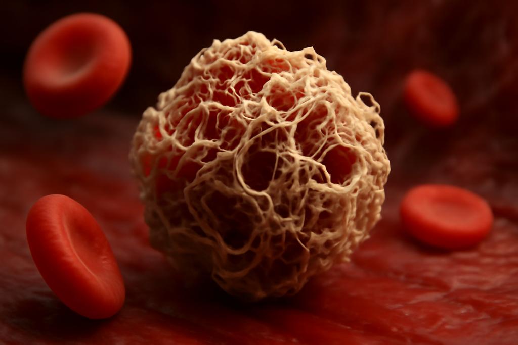
Blood clots are not static sculptures; they are living, bending mechanical systems that must yield to the river of blood while resisting it. The scaffold of a clot is fibrin, built from the protein fibrinogen, which is transformed into a fibrous network when the body needs to stop bleeding. In a study from Bennett University in Greater Noida, India, researchers Vivek Sharma and Poulomi Sadhukhan set out to explain how the tiny, folded domains inside fibrinogen decide how a clot stretches, twists, and recovers after being pulled by flow and pressure.
Their answer is a new kind of multiscale model that explicitly includes domain unfolding—the process by which small, folded protein regions open up and lengthen under force. The model, dubbed unfolding-incorporated coarse-grained polymer (UCGP), starts with a single fibrinogen monomer and scales up to a protofibril, a fiber, and finally a crosslinked network. It’s a way to connect molecular drama to the macroscopic properties we observe in clots inside living vessels. The goal isn’t to replace experiments but to build a bridge across scales so we can predict how a clot behaves under real-world stresses. The work is led by the authors at Bennett University, with Sharma and Sadhukhan steering the model toward the network level where health, disease, and therapy meet mechanics.
From monomer to protofibril
Fibrinogen is a three-domain star anchored by two long coiled-coil rods. In the model, each of the D and E globular domains is represented by a handful of beads connected by springs, while the coiled coils are a string of springs that hold the structure together. The unfolding of the D-domain—the region that anchors to its partner during assembly—happens in three steps. When the simulated force crosses a threshold, the model updates the spring constants and rest lengths to reflect unfolding, letting the D-domain grow from about 5 nanometers to as long as 80 nanometers, the E-domain from 1.5 to about 9 nanometers, and the coiled region from 17 to roughly 41 nanometers.
The team chose spring constants to mimic the relative stiffness of each region: a stiffer D-domain, a somewhat stiffer knob-like interaction, and a softer crosslinking region that ties protofibrils together later. As unfolding unfolds, the system reconfigures its internal springs so that the apparent length of the monomer swells, and the force needed to keep stretching shifts in characteristic steps. The model captures six unfolding events—three on each side of the monomer—producing a force-extension curve that dips each time a domain pops open and then resumes rising. It’s a fingerprint of unfolding leaving its mark on the macroscopic response, a sign that the protein’s hidden length is being revealed under stress.
This unfolding isn’t just cosmetic. The six dips align with the all-atom simulations that chart how a D-domain or a comparable module unfolds under force. The authors tune thresholds and rest lengths so the coarse-grained monomer, when stretched, mirrors the same sequence of events seen in those more detailed pictures. In a sense, the monomer behaves like a mechanical origami: it folds into a compact package and then, under enough pull, unfolds into longer, sleeker forms that alter the fabric of the fiber it will become.
Calibrating at the monomer scale is the crucial first step. It ensures that when the team climbs up to protofibrils and then fibers, they are not inventing new physics at every rung but carrying forward the same unfolding story. The monomer’s rules become the guidelines for how a protofibril behaves when many copies are jammed together and subsequently crosslinked into a network.
From protofibril to fiber and network
Protofibrils are short chains of fibrinogen units that assemble in a half-staggered arrangement through knob-hole interactions. In the model, this knob-hole binding is implemented with a very stiff, short spring, capturing how strongly those contacts lock the protofibrils in place. The D-domain near those contacts is modeled with a very short equilibrium length and high stiffness, reflecting how tightly the D-domain can compress space as the protofibril tightens and aligns during early polymerization. In parallel, the model keeps track of how the unfolded state of the D-domain influences the whole protofibril, because the lengthening of the monomer feeds into how the protofibril stacks and resists further stretching.
Fibers form when protofibrils run side by side and are crosslinked by the αC regions of the fibrinogen, represented as soft, long crosslinker springs. These crosslinkers are intentionally gentle in the model; their role is to couple adjacent protofibrils without turning the fiber into an impenetrable barricade. The researchers experimented with three scales of fiber: System1 with two protofibrils in parallel, System2 with five protofibrils, and System3 with fifteen protofibrils spanning a longer length. These “systems” serve as stepping stones from single molecules to thick fibers that resemble real fibrin strands in blood clots. In every case, the fiber is a parallel stack of protofibrils connected by crosslinkers, a structure that can bend, stretch, and unfold its internal domains as it is pulled apart.
To push the model toward a network, the authors build a two-dimensional triangulated mesh and assign a single spring to each edge of the triangulation that represents a segment of the largest fibrin fiber modeled. The initial spring length is about 5 nanometers, and the forward march of unfolding at the fiber level is triggered when the stress on a bond passes a threshold. Here the team introduces two important practical rules to keep the simulations sane at larger scales: only the bonds with the greatest extension unfold at any given moment, and only a fraction (about 10%) of eligible bonds will unfold in a single time step. This mirrors how real networks release stress gradually through many microscopic events rather than all at once in a dramatic, global flop.
Stretching the network under these rules reveals a remarkable cascade. When the monomer unfolds as a part of a fiber, the effective stiffness of that fiber drops briefly as length increases, and then rises again as more domains unfold and the network reorganizes. In larger networks, the unfolding events become less noisy and more patterned, yet the same four-part mechanical saga persists: an initial gentle rise (entropic elasticity), a steeper rise as the structure resists deformation (enthalpic elasticity), a plateau where unfolding relieves stress (a signature of the protein’s hidden length being revealed), and finally a renewed stiffening once most domains have unfolded. The model thus paints a consistent picture of how microscopic unfolding threads through macroscopic mechanics, even when scaled up to a network’s complexity.
What the model reveals about mechanics
One of the most striking outputs is the way the force-extension curves carve out distinct regimes. In a single protofibril or an isolated fiber, you see an initial entropic regime where the chain lengthens with little force, followed by an enthalpic regime where bonds and springs resist stretching. Then, as force climbs higher, unfolding of the mid-sized fibrin domains kicks in, creating a plateau: the system lengthens without a large increase in force because unfolding pockets are absorbing energy and lengthening the material. After the unfolding wave passes, the curve resumes a more linear rise with a new, higher slope reflecting the now-altered material. This bilinear-to-trilinear dance mirrors what experiments have long hinted at but rarely connected across scales: the nonlinear elasticity of fibrin fibers is not just a property of the network geometry but a direct fingerprint of domain unfolding inside the constituent proteins.
As the fiber grows from System1 to System3, fluctuations in the force-extension curves shrink. The same unfolding events occur, but the signals become cleaner as the system becomes larger and more redundant. Crosslinking—with its bridges between protofibrils—shifts the onset of stiffening and the height of the unfolding-related dips. When crosslinking is heavy, the network behaves more like a tight bundle; stress is distributed so more domains unfold in unison, and the first peak in the force-extension plot can be larger than the second. In contrast, with sparse crosslinking, the network acts more independently, and unfolding events are staggered, producing a different rhythm in the curve. These nuances matter because they hint at how real clots respond to the varying mechanical environments in vessels, where crosslink density and fiber organization can differ with tissue type, physiology, and disease.
Beyond fixed crosslinking, the researchers also explored the impact of alternative unfolding rules at the network level. When unfolding is allowed to cascade through an entire node (three-step unfolding) or is restricted to a single bond per step (one-step unfolding), the force-extension behavior shifts in characteristic ways. The three-step unfolding case yields a wide plateau and dramatic downstream softening, while the one-step case preserves a more bilinear signature. In one striking variant, diluted, selective unfolding at a node reproduces a bilinear response that echoes what has been observed in experimental studies of fibrin networks and gels. In short, the same molecular fiction—domain unfolding—can be told with several slightly different but plausible plotlines, depending on how stress is distributed and how many bonds are allowed to unlock at a time.
Why this matters for health, material design, and science
The practical upshot is that the primary engine behind the remarkable mechanical resilience of clots—the unfolding of globular domains inside fibrinogen—can now be studied in a scalable, multilevel way. The unfolding of the D-domain, the possible extension of coiled regions, and the subsequent rearrangements that unfold across protofibrils and across a network all contribute to a clot’s ability to stretch without breaking and then recover—an interplay that is central to clot stability in the bloodstream’s turbulent, pulsatile environment. This model provides a concrete framework to connect the microscopic logic of protein mechanics to the macroscopic signatures researchers measure in clots, gels, and networks. It is a rare bridge from atomistic detail to tissue-scale behavior that preserves the essential physics at each rung of the ladder.
Beyond understanding blood clots, the unfolding-incorporated coarse-grained polymer model opens a pathway for designing tougher, smarter biomaterials. Sacrificial bonds and domain unfolding are themes in tough, bio-inspired materials—bonds that give way under stress to absorb energy and then reform or rearrange. The UCGP approach shows how to tune the effective stiffness and the length gained from unfolding by adjusting a handful of parameters—how stiff a domain is, how long it unfolds, how readily crosslinkers hold fibers together. In other words, you could tailor a material’s mechanical response by choosing where and how much unfolding you want to happen, a design principle that could inform next-generation hydrogels, tissue scaffolds, or synthetic clots for medical applications.
From a broader science perspective, the work is a careful demonstration of multiscale modeling in a living system. It answers a central question the field has wrestled with: how do tiny molecular events ripple upward to alter the physics of a whole network? The authors emphasize that their model remains deliberately minimal—just enough to capture the key physics of domain unfolding, knob-hole interactions, crosslinking, and network topology—yet flexible enough to grow with future refinements. It’s a proof of concept that you can start from a single monomer, respect the microscopic drama inside, and still tell a coherent, testable story about the clot at the scale where doctors, patients, and machines live.
In the end, the unfolding story inside fibrinogen isn’t just about biology or physics; it’s a reminder that nature often solves tough problems by bending, lengthening, and redistributing stress. When a domain unfolds, it makes room for more stretch; when crosslinkers pull tighter, the same unfolding can be restrained; when the network yields a plateau instead of snapping, it buys time for recovery. These are the algebra and poetry of biology working in concert. The Bennett University study—led by Vivek Sharma and Poulomi Sadhukhan—provides a clear, testable map of that concert, offering a way to listen to the microscopic whispers that shape macroscopic strength and resilience in one of the body’s most vital, and most perilously delicate, structures.


