Framing the Magnetic Shadow Around MRI
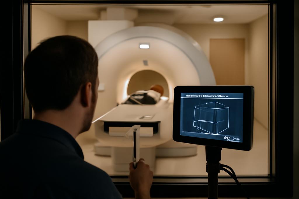
In every hospital MRI suite, a powerful magnet sits at the room’s heart, quietly shaping how patients are scanned and how researchers peer into the body. The magnetic field isn’t confined to the bore; it spills into the surrounding space, creating a magnetic landscape that human bodies and metallic objects can traverse. This fringe field, or stray field, is a familiar foe to MRI technicians and engineers: it can tug ferromagnetic objects toward the magnet, a danger popularly framed as the projectile effect. But there’s a second, subtler hazard that often travels under the headline: motion through the field can generate time-varying electric fields in tissues, which some people experience as headaches, dizziness, or the odd phosphenes in their vision.
What makes this study distinct is not just measuring how strong the field is, but mapping how it moves as you walk through the room. The 3T MRI era—where magnets push beyond 3 Tesla—has multiplied the need for precise, real-world maps of the magnetic environment. The better you can model the room’s magnetic geometry, the better you can predict what happens when a technician, a nurse, or a radiologist follows the real path they take during a typical scan. This isn’t just about avoiding accidents; it’s about building safety into the workflow itself, training staff with realistic scenarios, and designing spaces that minimize risk without slowing care.
These questions sit at the intersection of physics, medicine, and workplace safety. The study we’re looking at comes from Italian researchers who set out to create a comprehensive, three-dimensional map of the static magnetic field (B0) around a clinical 3T MRI system. It isn’t enough to know the field’s strength at a few points or along a couple of lines. To model motion accurately, you need a vector map: the magnitude |B| and the three axial components Bx, By, and Bz, throughout the room. That’s the heart of what the authors achieved, and it’s what makes the work both technically impressive and practically meaningful for people who spend their days in MRI suites.
How the 3D Map Was Built
The researchers’ approach is refreshingly pragmatic: they start with careful, unperturbed measurements of the magnetic field at a dense set of points in the room, focusing on areas where workers normally move. They used a commercial magnetometer, the HP-01, to capture not just the field’s magnitude but its full vector components. The measurements were collected on a grid that sits on planes parallel to the floor, at three distinct heights corresponding roughly to the anatomy of a person walking by—near the hips, chest, and head. In concrete terms, they laid down 252 measurement points across a frontal area of the room, then extended those measurements to reconstruct what the field looks like everywhere in the space.
But raw data in a real room is rarely neat enough to yield a full 3D map. So the team built a layered pipeline: first, quality control to prune outliers that sneak in because even a millimeter off can swing readings in such a sensitive environment. Then they fit the data with a family of nonlinear functions, choosing models that could capture both close-range, steep variations and long-range, slowly decaying trends. The fitting step involved comparing models by a reduced chi-squared metric to pick the best balance between bias and variance. After that, they filled in gaps with interpolation, using a natural-neighbor method that preserves physical continuity better than a lot of alternatives. Finally, they exploited the magnetic field’s symmetry to extend the mapping to all quadrants of the room, and they handled a small, special case around the magnet’s center where the field behaves like a 3 Tesla bubble.
Why all this rigour matters: the goal isn’t simply to produce a pretty picture. The researchers quantify uncertainty at every stage—instrument precision, fitting error, interpolation limitations—then combine these to estimate the total map’s reliability. In occupational exposure science, an acceptable total uncertainty hovers around 10 percent; the team’s analysis found a worst-case around 5.6 percent in the most challenging interpolated regions. That’s not a trivial accuracy bar; it’s the difference between a safe planning assumption and a risky guess when you’re predicting how a worker’s body might experience motion-induced fields.
Two hospital sites hosted the work, each with a different 3T MRI machine, testing the method’s generality. The first facility, FTGM Ospedale del Cuore in Massa, and the second, FTGM Ospedale San Cataldo in Pisa, provided real-world laboratories where shielding, architecture, and equipment interact with the field in unique ways. The result is a blueprint that isn’t tied to a single brand or room geometry but can be adapted to other clinical settings with a disciplined measurement plan and a principled interpolation toolkit.
From Data to a Living Room Map
One of the study’s striking moves is to treat the MRI room as a 3D volume, not a set of separate slices. With measurements on a 1-centimeter grid in the final 3D reconstruction, the authors produce a volumetric map of the magnetic field’s magnitude and, crucially, its axial components throughout the space. The endpoint is a digital twin of the room’s magnetic environment that can be interrogated for any position a worker might occupy. This is where the work shifts from an engineering curiosity into a practical tool for safety and training.
Central to the visualization is the isogauss surface concept—the sheets of equal magnetic field strength that sweep through the room. The team didn’t stop at exposing regions where |B| exceeds a threshold; they also mapped the directional field lines and the vector components that determine how rapid movement in the room can induce electric fields in tissues. A spherical region, about 60 centimeters in diameter, around the isocenter remains at a constant 3 Tesla, reflecting the core magnetic core of the scanner. Surrounding this core, the fringe field fades and twists in complex patterns shaped by the room’s geometry and the scanner’s shielding. Mapping all of this with high fidelity is what lets you answer practical questions such as: where should a nurse avoid a certain stretch of floor, or how might a particular patient’s head move through a rotating field?
The authors emphasize that the method is not a compliance checklist but a simulation-enabling tool. With a faithful 3D map, digital models can run trajectories—the paths a technician might trace during a standard workflow—and compute the resulting induced electric fields in the body. That, in turn, informs whether a given movement would push exposure toward regulatory limits or simply make a worker feel unwell for a moment. It’s a shift from static safety margins to trajectory-aware risk assessment, which aligns with how modern workplaces actually operate: people move, objects move, and the room is a dynamic stage rather than a still background.
Why This Changes MRI Safety Practice
Safety culture in MRI suites has long rested on three pillars: standardized rules, device-specific guidelines, and the implicit trust that the room’s physical layout is well understood. What this study adds is a way to quantify and visualize what happens as people actually move through the space. It’s the difference between a map that shows where the danger lives and a map that shows how danger travels as you walk, lean, twist, or pivot. The practical upshot is twofold: better training and better workflow design.
Training becomes more realistic when staff can watch, in a simulated map, how their typical movements push them into higher-gradient zones or into parts of the room where the axial components of the field matter more for rotation or torsion. Trainees can experiment with different paths and speeds, seeing how small choices—where to stand, how to turn, when to step back—affect exposure. For operators, the 3D map offers a shared language for planning layouts that minimize dangerous trajectories, or for selecting equipment placement so that critical tasks stay within safer corners of the room. In essence, the map translates abstract physics into actionable, embodied knowledge.
Another major implication lies in the potential to design more intelligent training and safety protocols. The digital map can feed into simulations that estimate exposure for various job scenarios, helping institutions define zone boundaries, equipment placement, and standard operating procedures that account for motion-induced fields. The authors suggest that their approach can evolve into an educational tool—one that teaches operators not just where a red zone is, but how to navigate the room in ways that keep electric-field exposure as low as reasonably achievable during routine work.
Beyond Static Fields: Moving Bodies in a Static World
One of the most provocative ideas in the paper is the explicit attention to axial components, not just the field’s magnitude. In practical terms, when a person turns their head, twists their torso, or tilts their body, different components of the magnetic field interact with the body differently. The axial components (Bx, By, Bz) determine the induced electric fields that arise from a given motion via Faraday’s law. If you only know |B|, you miss a lot of the story about what happens when people move in three dimensions. This is especially important for rotational movements—the kinds of motions that happen naturally as clinicians examine a patient or adjust a coil, head support, or positioning aids.
And there’s a cautionary note for the push toward ever-stronger scanners. As magnetic fields get stronger, fringe fields don’t simply recede; they morph in ways that require more careful modeling. The paper points out that newer scanners deploy active shielding and other architectural features that alter the field’s exterior profile in nontrivial ways. If you rely on simplified dipole approximations or on manufacturer-isogauss maps that emphasize magnitude but ignore direction, you risk underestimating or mischaracterizing exposure, particularly for complex movements. The 3D vector map offers a more faithful canvas for simulating real-world exposures as mobile workflows evolve and MRI technologies advance.
In short, the study reframes a familiar safety problem as a data-rich, geometry-aware challenge. It invites a future where every MRI room has a digitally mapped magnetic personality, one that can be interrogated to answer: how does a specific sequence of moves translate into physical sensations or potential risks? That accountability is valuable not just for worker safety but for the design of future MRI facilities themselves.
The Human Angle: Who Built the Map and Why It Matters
Behind this work stands a collaboration among Italian researchers anchored in three institutions: the Institute of Clinical Physiology, CNR (National Research Council) in Pisa, the Department of Occupational and Environmental Medicine at INAIL in Rome, and the Department of Biomedical, Dental, and Image Sciences at the University of Messina. The paper’s authors include Francesco Girardello, Maria Antonietta D’Avanzo, Massimo Mattozzi, Victorian Michele Ferro, Giuseppe Acri, and Valentina Hartwig, among others. The lead voices—Valentina Hartwig and Giuseppe Acri—are cited repeatedly for conceptualizing the mapping approach and driving the analysis that turns scattered field measurements into a coherent 3D model.
What makes this collaborative effort meaningful is its dedication to real clinical environments. The researchers tested their pipeline in two hospital facilities with 3T scanners from different manufacturers, a deliberate choice that demonstrates how the method can adapt across machines, room geometries, and shielding configurations. It’s a reminder that safety solutions in high-tech medicine don’t live in a lab alone; they must travel into real rooms where patients lie, technicians pace, and the everyday choreography of care unfolds. The study reads like a blueprint for institutions that want to elevate their MRI safety programs from reactive compliance to proactive, data-informed design.
As with any work that seeks to quantify the invisible, there are caveats. The map is as good as the measurements it rests on, and even with careful interpolation, some regions—especially those inside the central magnet where direct axial data can’t be measured—remain more uncertain. The authors are transparent about these limits, framing the map as a tool for education, planning, and risk estimation rather than a universal, one-size-fits-all safety certificate. They clear-eyedly propose that the map’s true power lies in its use as a dynamic, experiment-ready platform: plug in a movement trajectory, run a simulation, and watch how exposure evolves in time and space.
One of the study’s most human insights is its implicit invitation: to rethink how we train MRI teams for a future in which technology is steadily stronger and workspace designs shift accordingly. The authors are not arguing for more bureaucracy; they’re offering a practical asset—an accessible, precise, and adaptable map—that can empower staff to navigate the room with greater awareness, confidence, and, ultimately, safety. If safety is a culture, this map helps seed a culture where every movement is accounted for, every trajectory anticipated, and every staff member equipped with a better understanding of the magnetic world they inhabit daily.
The People Behind the Map and the Road Ahead
The work is a vivid reminder that scientific progress in clinical settings is rarely a solo act. It’s the fruit of multidisciplinary teamwork—physics, occupational health, medical imaging, and engineering—applied to real patients and real staff in real rooms. The institutions involved—Instituto di Fisiologia Clinica at CNR in Pisa, INAIL in Rome, and the University of Messina—each brought their strengths to the table: rigorous measurement, robust exposure assessment, and a clinical-eye for how safety translates into everyday practice. The study demonstrates what happens when researchers move beyond theory to test methods in actual hospital environments, with the variability and constraints those environments inevitably present.
Looking forward, the authors envision a family of extensions. They hint at using the 3D maps to estimate exposure for specific body parts during a broader set of movements—displacement, rotation, flexion—so that more granular, trajectory-based exposure assessments become feasible. There’s also a natural path to apply the methodology to different field strengths and scanner designs, expanding the library of rooms that can be modeled with the same disciplined protocol. In a healthcare world where MRI technology keeps getting sharper and the demand for safer, more efficient care grows, this kind of forward-looking, practically grounded research could become a staple of how hospitals design, operate, and train for safety around powerful magnets.
In the end, the study offers a quiet kind of revelation: there are invisible forces at play in MRI rooms, but with careful measurement, thoughtful modeling, and a commitment to translating science into practice, we can illuminate those forces well enough to protect the people who navigate them every day. The map is not just a scientific curiosity; it’s a tool for safer care, a teaching aid for staff, and a stepping-stone toward smarter, trajectory-aware safety in a future where MRI machines continue to push the boundaries of what medicine can do.
Takeaway: by turning the fringe field into a full 3D vector map, researchers provide a practical bridge between high-field physics and everyday safety—helping MRI teams move through their magnetic landscape with greater awareness, better training, and a clearer path toward safer, more effective imaging.


