Unlocking Genetic Clues from Ordinary Tissue Slides
In the fight against lung cancer, knowing the enemy’s genetic makeup can be the difference between life and death. Certain mutations in cancer cells—called driver mutations—act like switches that fuel tumor growth. Targeted therapies that shut off these switches have revolutionized treatment, turning what was once a grim prognosis into a manageable condition for many patients. But here’s the catch: identifying these mutations requires genetic testing, a process that’s often expensive, slow, and inaccessible in many hospitals around the world.
Enter a team of researchers from Lunit Oncology, a cutting-edge medical AI company with labs in Germany and South Korea, who have developed a clever machine learning approach that reads these genetic secrets not from DNA sequencing, but from the humble microscope slides pathologists use every day. Their work, led by Biagio Brattoli and Jack Shi, introduces a new model called the Asymmetric Transformer Decoder, which can predict six key lung cancer driver mutations by analyzing routine stained tissue images.
Why This Matters More Than Ever
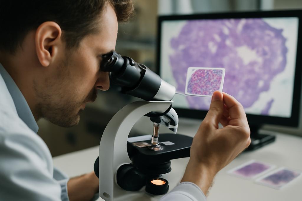
Genetic testing for lung cancer mutations like EGFR, KRAS, ALK, and others is recommended worldwide because it guides doctors to the most effective treatments. But the reality is that many patients never get tested due to cost, lack of specialized labs, or delays that push back critical therapy decisions. Meanwhile, histology slides—thin slices of tumor tissue stained with hematoxylin and eosin (H&E)—are available everywhere. These slides contain subtle visual clues about the tumor’s biology, but interpreting them to infer genetic mutations is a task beyond human eyes.
Machine learning models have shown promise in bridging this gap, but previous efforts mostly focused on one or two mutations, limiting their clinical impact. The Lunit team’s approach is broader and smarter: it simultaneously predicts six actionable mutations, including some that are rare and notoriously difficult to detect.
Decoding Complexity with an Asymmetric Transformer
Whole slide images (WSIs) are enormous—imagine gigapixel photos of tissue sections. Feeding these directly into a computer model is like trying to swallow an elephant whole. The standard workaround is to chop the image into thousands of smaller patches and analyze them piece by piece. But then the challenge becomes how to combine all these patch-level insights into a coherent prediction about the entire slide.
This is where Multiple Instance Learning (MIL) comes in. MIL treats each slide as a “bag” of patches, learning to predict slide-level labels without needing patch-level annotations. However, not all MIL methods are created equal. The Lunit researchers found that conventional transformer models, which have taken the AI world by storm, struggle when applied naively to this problem. Transformers typically expect queries, keys, and values to have the same dimensions, but this can lead to bloated models prone to overfitting—especially when data is limited.
Their solution? An Asymmetric Transformer Decoder that cleverly uses queries and key-value pairs of different sizes. Think of it as a translator who listens carefully to a complex story (high-dimensional patch features) but summarizes it succinctly (low-dimensional queries) to avoid getting lost in the noise. This design lets the model extract rich information efficiently without drowning in irrelevant details.
Listening to the Tumor’s Neighborhood
Another innovation is how the model respects the biological context of each patch. Tumors don’t exist in isolation; they interact with surrounding stroma (supportive tissue) and even seemingly normal background areas. Previous models often focused only on cancerous regions, potentially missing important signals.
The Lunit team used a segmentation model to classify patches into three tissue types: cancer area, cancer stroma, and background. They then embedded this tissue-type information directly into the model’s input, allowing it to weigh each patch’s contribution based on its biological role. This approach is like giving the model a map of the tumor’s neighborhood, helping it understand not just what it sees, but where it sees it.
Results That Inspire Hope
Testing their model on over 1,200 lung cancer slides from diverse hospitals, the researchers demonstrated that their Asymmetric Transformer Decoder outperformed state-of-the-art MIL methods by an average of 3%, and by more than 4% when detecting rare mutations like ERBB2 and BRAF. These improvements might seem modest at first glance, but in the high-stakes world of cancer diagnosis, every percentage point can translate into better treatment decisions for thousands of patients.
Moreover, when validated on an external dataset from The Cancer Genome Atlas, the model maintained strong performance, underscoring its potential generalizability across different populations and scanning technologies.
Bringing Precision Oncology to More Patients
The implications of this work are profound. By harnessing existing pathology workflows and standard H&E slides, this AI-driven approach could democratize access to genetic insights, especially in resource-limited settings where sequencing is not feasible. It could serve as a rapid screening tool to identify patients unlikely to harbor certain mutations, sparing them unnecessary delays and costs, while flagging those who should undergo confirmatory genetic testing.
In a way, this model acts like a seasoned detective, piecing together subtle visual clues from the tumor’s microenvironment to reveal its hidden genetic identity. It’s a testament to how artificial intelligence, when thoughtfully designed and biologically informed, can amplify human expertise and bring personalized medicine within reach for more people.
Looking Ahead
While promising, this technology is not a replacement for genetic testing—yet. Further clinical validation, integration into diagnostic workflows, and regulatory approvals lie ahead. But the study from Lunit Oncology offers a glimpse of a future where the microscope and the computer work hand in hand, turning routine pathology slides into powerful windows into cancer’s genetic code.
In the relentless quest to outsmart cancer, sometimes the answers are hidden in plain sight, waiting for the right lens to bring them into focus.


