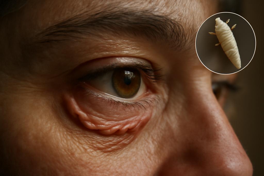
Imagine a world where eye exams could reveal hidden diseases, not just through what your doctor sees, but through intricate patterns invisible to the naked eye. That future might be closer than we think, thanks to a groundbreaking study by researchers at Zhejiang Normal University and Wenzhou Medical University, led by Shoujun Huang and Qi Dai. Their work unveils a novel method for analyzing the subtle twists and turns within medical images, offering a potentially transformative approach to diagnosis.
The Winding Road to Diagnosis
The research focuses on something called “tortuosity.” In essence, it’s the measure of how curvy a line is. Think of a river snaking through a landscape versus a straight canal. The river is tortuous; the canal, not so much. This concept becomes incredibly relevant in medical imaging, where the shapes of blood vessels, nerves, and even glands often hold crucial clues to underlying health conditions.
Existing methods for measuring tortuosity often rely on simplistic comparisons to a perfectly straight line. But as the researchers point out, many biological structures have a natural, healthy amount of curve. Using a straight line as a benchmark can be like judging a sculptor’s work by how closely it resembles a block of untouched marble. It misses the nuances of the actual form and what makes it meaningful.
A New Map: Information Entropy and Curve Comparison
The team at Zhejiang Normal University and Wenzhou Medical University devised a more sophisticated approach. Their innovative framework uses something called “information entropy,” a concept borrowed from information theory. Entropy essentially quantifies disorder or randomness in a system. By comparing a target curve (e.g., the outline of a meibomian gland in an eye image) to a reference curve (a healthy, less tortuous version of the same structure), they can measure how much the target curve deviates from the norm – and thus, how much “disorder” or irregularity it displays.
The beauty of this method lies in its adaptability. It doesn’t just compare against an idealized straight line; it allows for a more realistic, biologically plausible comparison. This is crucial when dealing with complex biological structures, like meibomian glands, whose healthy shapes aren’t perfectly straight but have their own natural curves.
Unmasking Demodex: A Clinical Application
The researchers tested their method on images of meibomian glands, tiny oil-producing glands in the eyelids. Infections by Demodex mites are often associated with uneven atrophy of these glands, potentially contributing to dry eye disease. This unevenness would be reflected in the tortuosity of the glands’ outlines in images. The new approach aimed to accurately and quantitatively assess this irregularity.
Their analysis of 105 patients showed remarkable success. The information entropy-based framework distinguished between patients with Demodex infections and those without with impressive accuracy. The results achieved an area under the curve (AUC) of 0.858 in receiver operating characteristic (ROC) analysis, with a sensitivity of 0.786 and a specificity of 0.857 – far better than traditional methods. This suggests that the method could offer a fast, objective, and cost-effective diagnostic tool to replace subjective clinical assessment.
Beyond the Eye: Wider Implications
The implications of this study extend far beyond the diagnosis of Demodex infections. The information entropy-based framework is a generalizable method applicable to analyzing the tortuosity of various structures in medical images. Think about blood vessels in the brain, damaged nerves in the cornea, or even the winding paths of intestinal tracts. This technique could help detect abnormalities in a host of diseases and conditions.
Of course, the approach isn’t without its limitations. The choice of reference curve, for instance, is critical for the accuracy of the assessment. And while the method successfully distinguishes between healthy and diseased states in their test case, more extensive validation is needed. Future work will focus on refining the standard curve selection process and addressing computational complexities to enable more widespread and practical clinical use.
The Human Element in Algorithmic Precision
What’s truly exciting about this research is the way it combines sophisticated mathematical tools with the subtle visual cues of medical imaging. The team’s approach is not a mere replacement for human expertise. Instead, it’s a powerful augmentation. This method doesn’t eliminate the doctor; it enhances their capabilities, enabling more precise diagnoses and potentially life-saving interventions. It is a testament to the power of interdisciplinary collaboration, blending the rigorous logic of mathematics and computer science with the profound understanding of human biology and disease.
This technology is a powerful example of how seemingly abstract concepts in mathematics and engineering can translate into practical medical tools that ultimately improve human lives. It is a fascinating glimpse into a future where data analysis helps uncover hidden information within medical images, leading to earlier, more accurate, and more effective healthcare.


